

Imagine finding a painless lump on the side of your neck. You are not in pain. You feel fine. But the lump has been there for a year, maybe longer, quietly growing. Or perhaps you have developed a rhythmic whooshing sound in one ear, perfectly in time with your heartbeat. These are two of the most common ways people first encounter a head and neck paraganglioma: a rare, slow-growing tumour that most people have never heard of.
This article explains what these tumours are, where they occur, how doctors find them, what treatment involves, and what you can expect in terms of outcomes. Because up to 40% of cases have a genetic cause, we also cover what that means for you and your family.
What Exactly Is a Paraganglioma?
Your body contains clusters of specialised nerve cells scattered along major blood vessels and nerve pathways. These clusters, called paraganglia, originally developed during foetal life from a type of cell known as neural crest cells. Their job is to help the body sense and respond to changes in blood pressure, oxygen levels, and other physiological signals.
A paraganglioma is a tumour that grows from one of these clusters. When one forms in the head or neck region, it is called a head and neck paraganglioma, often abbreviated to HNPGL. You may also hear them called "glomus tumours," named after their most common locations: the glomus jugulare (near the base of the skull), the glomus tympanicum (in the middle ear), and the carotid body (in the neck).
The overwhelming majority of these tumours are benign: they do not invade other organs or spread through the bloodstream. They do, however, grow in tricky anatomical territory, wrapped around arteries and nestled against nerves that control your voice, swallowing, hearing, and shoulder movement. That is what makes them clinically significant despite their usually non-cancerous nature.
How Common Are They?
These are genuinely rare tumours. The annual incidence is roughly 0.3 to 1 case per 100,000 people, and temporal bone paragangliomas (the middle-ear type) occur in only about 1 per 1.4 million people per year. Despite this rarity, the incidence appears to be rising, largely because modern CT and MRI scans detect tumours that would previously have gone undetected for years.
Head and neck paragangliomas account for approximately 65–70% of all paragangliomas found in the human body, making the head and neck the single most common region affected. They account for less than 1% of all head and neck tumours.
The typical age at diagnosis for sporadic (non-inherited) cases is the fourth to fifth decade of life, with a range from 15 to 85 years. Women are diagnosed more frequently than men overall, particularly with glomus jugulare and tympanicum tumours, where the female-to-male ratio can reach 4 to 6:1. Hereditary cases tend to appear about a decade earlier in life.
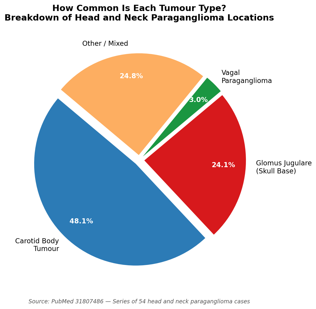 Figure 1: Distribution of head and neck paraganglioma subtypes. The carotid body is the most common site, accounting for approximately 48% of cases—data from peer-reviewed institutional series.
Where Do These Tumours Grow?
There are four main locations, each producing a distinct set of symptoms and surgical challenges.
Carotid body paraganglioma is the most common type, accounting for roughly 48% of all head and neck paragangliomas. The carotid body sits at the fork where the main artery of the neck divides into its two branches. A tumour here typically presents as a painless, slowly enlarging lump on the side of the neck. As it grows, it pushes these two artery branches apart, a characteristic sign that shows clearly on imaging. If it grows large enough to press on nearby nerves, it can cause hoarseness or difficulty swallowing.
Jugular paraganglioma (glomus jugulare) accounts for around 24% of cases. This tumour grows deep inside the skull base, at the point where the jugular vein exits the skull through a channel surrounded by several important cranial nerves. Because of its location, it tends to produce pulsatile tinnitus (a heartbeat-like sound in one ear), hearing loss, and nerve problems affecting swallowing, voice, and shoulder strength.
Tympanic paraganglioma (glomus tympanicum) grows on the inner wall of the middle ear. It is the most common tumour of the middle ear overall. The classic presentation is pulsatile tinnitus and conductive hearing loss, and an examining doctor may be able to see a characteristic red mass through the eardrum using an otoscope. Hearing loss is reported in roughly 80% of patients with temporal bone paragangliomas, and pulsatile tinnitus in about 77%.
Vagal paraganglioma is the rarest type, accounting for only around 3% of all head and neck paragangliomas. It arises from nerve clusters within the vagus nerve, which runs down the neck, controlling voice, swallowing, and even heart rate. It usually presents as a painless mass in the upper neck or behind the jaw, sometimes accompanied by hoarseness or a cough triggered by touching the neck.
Recognising the Symptoms
Because these tumours grow so slowly, there is often a gap of four to seven years between the first symptoms and a firm diagnosis. Many people assume a slow-growing neck lump is a lymph node, or attribute ear symptoms to other causes. The routes to diagnosis typically include:
- A painless, slowly enlarging lump in the side of the neck
- Pulsatile tinnitus: a rhythmic whooshing in one ear, timed with the heartbeat
- Unexplained hearing loss, particularly one-sided
- Hoarseness, voice changes, or difficulty swallowing
- A red or blue mass visible through the eardrum on examination
- Incidental discovery on a CT or MRI scan done for an unrelated reason (this accounts for roughly 25–30% of cases)
- Proactive genetic screening in people known to carry a paraganglioma-associated gene variant
About 9% of head and neck paragangliomas secrete hormones (catecholamines, the adrenaline family). When this happens, patients may experience episodic headaches, rapid heartbeat, sweating, and spikes in blood pressure. These symptoms can mimic anxiety or thyroid disease, which sometimes leads to delays in diagnosis. Biochemical testing (measuring metanephrines in blood or urine) is carried out in all newly diagnosed patients to check for this, since secreting tumours need special preparation before any surgical procedure.
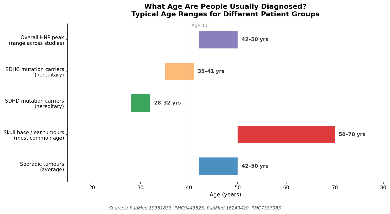 Figure 2: Age at diagnosis distribution. Peak incidence falls in the fourth and fifth decades of life for sporadic cases; hereditary cases tend to present roughly ten years earlier. Data from multiple published cohort studies.
How Doctors Find and Confirm the Diagnosis
Diagnosing a paraganglioma typically involves several layers of investigation, moving from initial suspicion to detailed mapping before treatment.
Ultrasound is usually the first test for a neck lump. It reveals a highly vascular (blood-rich) mass and can point strongly toward the diagnosis, but it cannot image tumours deep within the skull base.
CT scanning with contrast provides excellent detail of bone anatomy, shows exactly how the tumour relates to surrounding blood vessels, and reveals bone erosion in jugular tumours. On CT angiography, paragangliomas show a striking "lightbulb sign": they light up very brightly and early with contrast dye because of their exceptionally rich blood supply. CT angiography also demonstrates the characteristic splaying of the carotid artery branches in carotid body tumours.
MRI with gadolinium contrast gives superior detail of soft tissues, nerves, and intracranial (inside-the-skull) extension. On certain MRI sequences, paragangliomas display a classic "salt and pepper" appearance, produced by a mix of bright spots where blood flows slowly and dark signal voids where blood flows rapidly through the tumour's many vessels. Contrast-enhanced MR angiography achieves a sensitivity of 100% and specificity of 94% for detecting these tumours.
Digital subtraction angiography (DSA) maps the exact blood supply feeding the tumour by injecting contrast dye directly into the arteries via a catheter. This is performed when surgery is planned, because paragangliomas are notoriously difficult to remove.
68Ga-DOTATATE PET/CT is the most advanced imaging tool for paragangliomas. It uses a radioactive tracer that binds specifically to receptors found on paraganglioma cells, allowing a single whole-body scan to detect every tumour site simultaneously. Detection rates per lesion reach 92–100%, with a specificity of 96.6%. This matters enormously because 10–20% of patients harbour more than one tumour at the time of diagnosis. This scan is now recommended as the first-line staging investigation for known or suspected paraganglioma.
Treatment Options: Surgery, Radiation, or Watch and Wait?
There is no single right answer for every patient. Treatment decisions depend on the tumour's location, size, relationship to blood vessels, your age and general health, and whether you carry a genetic mutation associated with higher malignancy risk. Three main strategies exist.
Surgery
Surgery is the traditional gold standard and remains the preferred approach for most carotid body tumours and for younger, otherwise healthy patients with accessible tumours. In experienced specialist centres, complete surgical removal is achieved in 92–100% of carotid body cases. After complete resection of jugular paragangliomas, the cure rate approaches 90%.
The main risk of surgery is injury to the cranial nerves that pass through or near the tumour. For carotid body tumours, surgeons use the Shamblin classification to predict this risk, grading tumours from Class I (minimal contact with the artery) through Class III (completely encasing the artery). Class III tumours carry a cranial nerve deficit rate as high as 83% immediately after surgery, though many of these deficits improve with time.
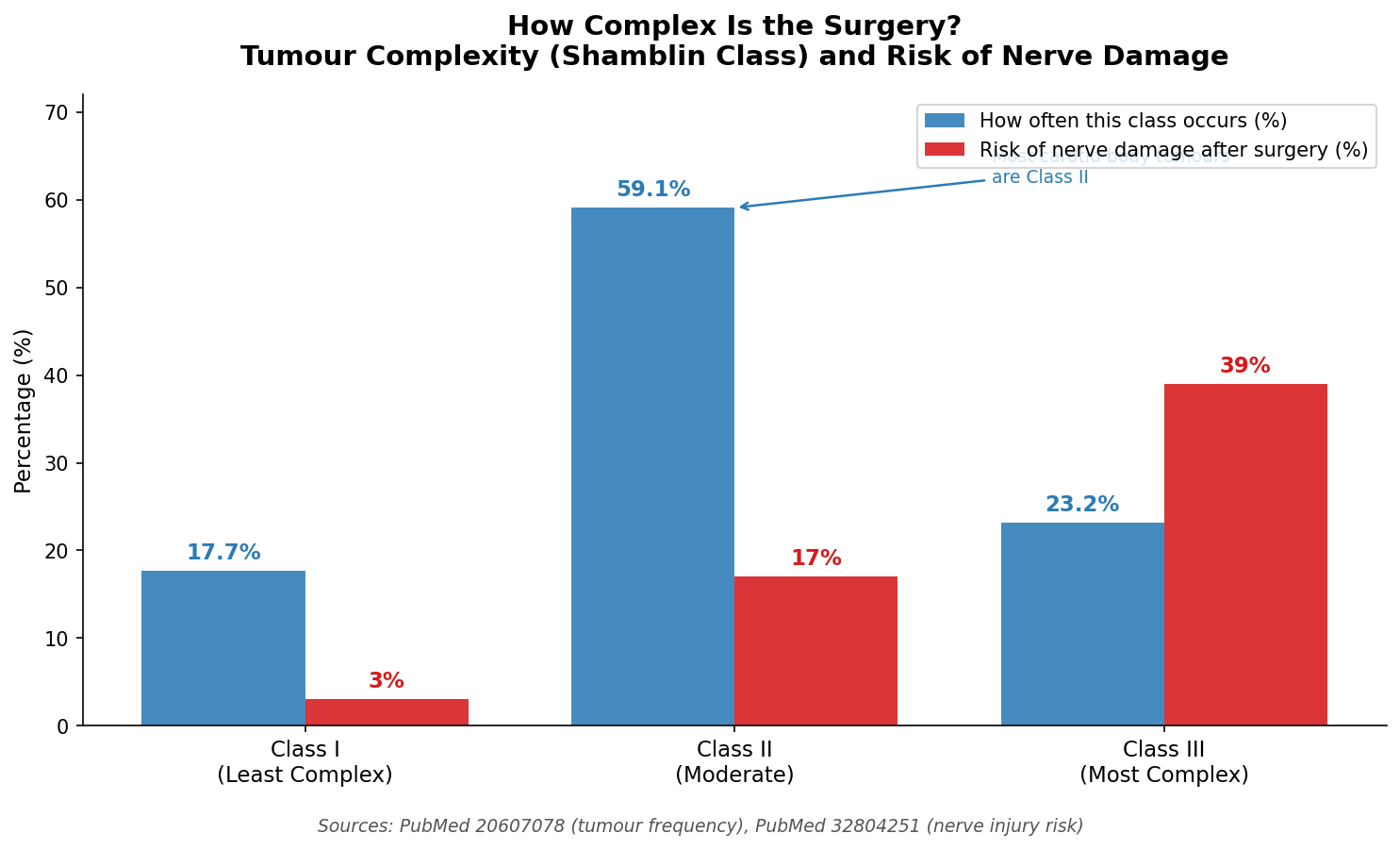 Figure 3: Shamblin classification system for carotid body paragangliomas, showing surgical complexity and associated cranial nerve injury risk by class. Data from published surgical series (PubMed 20607078, 32804251)
For jugular and vagal paragangliomas, permanent cranial nerve damage is considerably more likely: roughly 45% of patients undergoing surgery for jugular tumours experience some degree of permanent nerve deficit. Among patients in one large series, 13% required a feeding tube after surgery, 23% needed a procedure to improve voice function, and 3.3% suffered a stroke.
Preoperative embolisation is a procedure performed 24–48 hours before surgery in many centres. A radiologist threads a catheter into the arteries supplying the tumour and blocks them, dramatically reducing the blood supply. Studies show this cuts intraoperative blood loss by approximately 57% (from a mean of 1,374 mL without embolisation to 588 mL with it) and shortens operating time by roughly an hour.
Radiation Therapy
Radiation has become an increasingly preferred treatment, particularly for jugular and vagal paragangliomas, where surgery carries a high risk of nerve damage. Modern radiation techniques include conventional external beam radiotherapy (EBRT), intensity-modulated radiotherapy (IMRT), and stereotactic radiosurgery (SRS), where a single high-dose session is delivered to the tumour with sub-millimetre precision using systems such as the Gamma Knife or CyberKnife.
Radiation does not usually eradicate the tumour cells outright; instead, it halts growth and achieves long-term control. Published studies show remarkable outcomes:
- A 45-year EBRT experience: 99% five-year local control, 96% at ten years
- A 35-year series: 96% five-year control, 94% at ten years, 90% at fifteen years
- Gamma Knife radiosurgery as primary treatment for jugular paragangliomas: 100% progression-free survival at five, ten, and fifteen years
- A 23-year IMRT retrospective: 100% local control at both five and ten years, with zero cases of significant acute or late toxicity
Critically, radiation preserves the cranial nerves far better than surgery. A study comparing radiosurgery-treated jugular paraganglioma patients with general population norms found that quality-of-life scores (physical and mental) were similar to national averages, in sharp contrast to surgical series, where permanent nerve deficits substantially affected daily function.
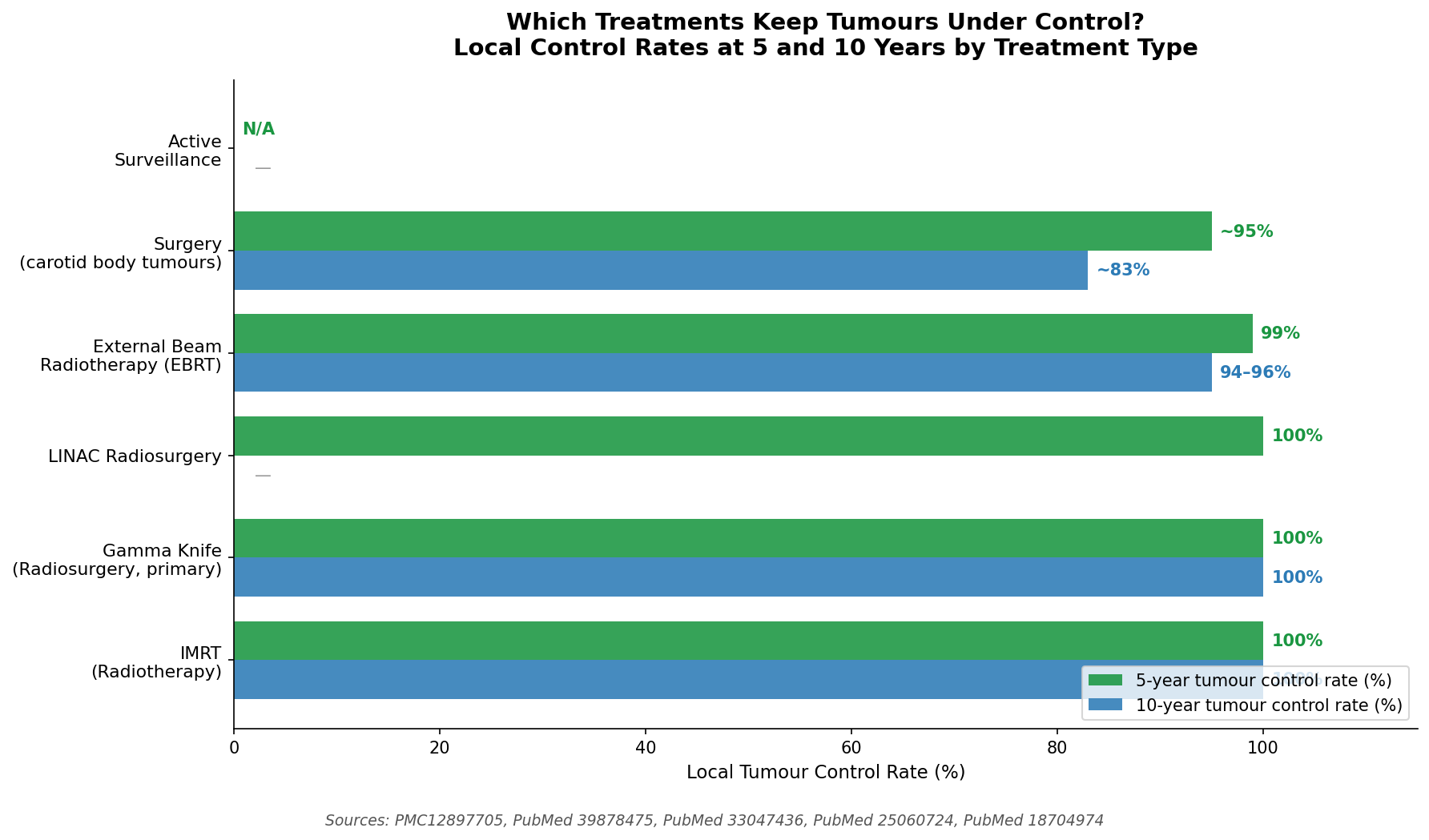 Figure 4: Local tumour control rates across treatment modalities. Surgery and radiation achieve broadly similar long-term control, but radiation carries substantially lower rates of cranial nerve complications for skull base tumours. Data from multiple published series.
Active Surveillance ("Watch and Scan")
For older patients, those with multiple serious health conditions, or those with small, asymptomatic tumours found incidentally, active surveillance is a legitimate and well-supported strategy. This means regular MRI scans (typically every one to two years) to monitor for any growth, without immediate treatment.
This approach is justified by the biology of these tumours. The median growth rate is only 1.0 mm per year, and the median time for a tumour to double in volume is 4.2 years. In natural history studies, approximately 42% of observed tumours showed no measurable growth at all during follow-up. Crucially, patients managed with observation alone consistently avoid the cranial nerve problems that affect a significant proportion of those who undergo surgery.
The key proviso is that observation is not "do nothing." It requires lifelong commitment to regular imaging, with a plan to escalate to treatment if the tumour grows, begins to cause symptoms, or shows features of aggressive behaviour.
The Surgical Decision in More Detail
For surgeons and their patients, the Shamblin class of a carotid body tumour is the single most important pre-operative factor. A Class I tumour sits mostly beside the artery and can be peeled off relatively safely. A Class III tumour fully encases the carotid artery, sometimes requiring vascular surgeons on standby who are prepared to reconstruct the artery if it is damaged or needs to be resected.
For skull base tumours (jugular and vagal types), the Fisch classification grades tumours from A to D based on how far they have grown toward the brain and surrounding structures. Higher Fisch grades are associated with far greater surgical complexity and nerve risk, which is a primary reason why many skull base centres now favour radiation as first-line treatment for these locations.
Complications to be aware of include:
- Cranial nerve deficits affecting voice, swallowing, shoulder function, or tongue movement
- Stroke (approximately 3% in complex surgical series)
- Blood loss, particularly in larger Shamblin II and III tumours without preoperative embolisation
- Very rarely, operative death (reported at 0% in major specialist series, reflecting the importance of case volume and expertise)
What Happens After Treatment: Recurrence and Long-Term Monitoring
One of the most important facts about paragangliomas is that recurrence can happen many years after seemingly successful treatment. Overall, published recurrence rates are 8.2% at four years and 17.1% at ten years across all treatment modalities. The mean interval from initial treatment to recurrence is 9.7 years, and a third of all recurrences appear more than ten years after treatment.
This has a direct and practical implication: lifelong surveillance is mandatory. Unlike many cancers, where "ten years all clear" is considered a cure, paraganglioma patients require imaging for the rest of their lives.
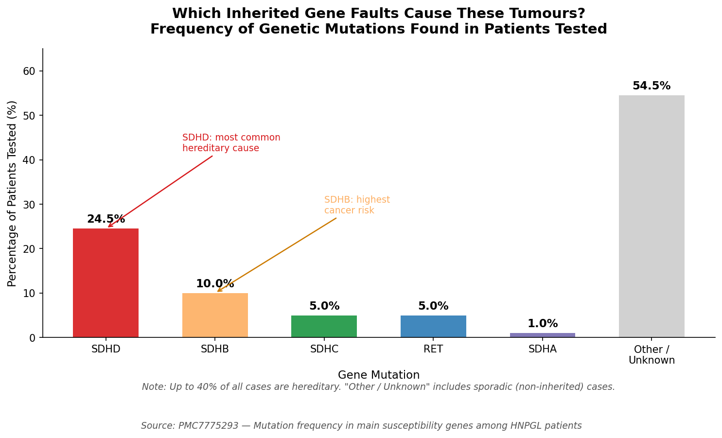 Figure 6: Recurrence rates over time following treatment. The long tail of recurrences beyond ten years underscores the need for lifelong surveillance imaging. Data from published follow-up studies)
Radiation therapy is associated with lower recurrence risk compared to surgery in most comparative studies. Gamma Knife radiosurgery for jugular paragangliomas as primary treatment showed 0% recurrence at five, ten, and fifteen years in one large specialist series, though all patients still require regular scanning.
Outcomes and Prognosis
For the majority of patients whose paraganglioma is benign, which is the case for around 85–90% of people, the long-term outlook is excellent. Tumour control rates with modern surgery or radiation exceed 90% in specialist centres. Quality of life after radiation therapy is broadly comparable to that of the general population.
The small minority (6–19% depending on the study) whose tumours are malignant (confirmed by spread to lymph nodes or distant organs) face a more complex picture:
- Five-year relative survival (all malignant head and neck paragangliomas): 59.5%
- Five-year survival for disease confined to regional lymph nodes: 76.8%
- Five-year survival with distant spread: 11.8%
- Five-year survival after surgery alone for malignant tumours: 88.1%
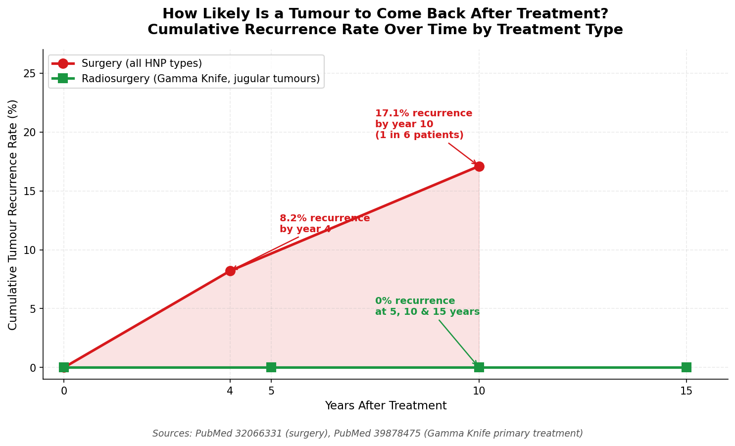 Figure 7: Five-year survival rates for malignant head and neck paragangliomas by stage of disease. Outcomes are substantially better when the disease remains regionally confined. Data from the National Cancer Database.
Malignancy is defined not by how a tumour looks under a microscope (because benign and malignant paragangliomas can look identical on pathology) but by whether it has spread. The SDHB genetic mutation carries the highest malignancy risk: roughly 31% of patients with this mutation develop metastatic disease, compared with a much lower rate in other gene variants.
Genetics, Inheritance, and Family Screening
Perhaps the most distinctive feature of paragangliomas among all tumour types is their strong inherited component. Up to 40% of all head and neck paragangliomas are caused by an inherited mutation in one of several susceptibility genes, with mutations in the succinate dehydrogenase (SDH) gene complex being the most important.
Think of the SDH genes as part of the cell's energy-production machinery. When they malfunction due to an inherited mutation, they can trigger abnormal growth in paraganglia cells over time. Nine susceptibility genes have been identified: SDHD, SDHB, SDHC, SDHAF2, SDHA, VHL, RET, NF1, and TMEM127.
The most clinically relevant:
- SDHD is the most common mutation found in head and neck paragangliomas (around 24.5% of tested patients). It is particularly associated with multiple tumours: up to 54% of SDHD carriers eventually develop more than one paraganglioma. Tumours tend to appear around age 30
- SDHB carries the highest malignancy risk. About 31% of carriers develop metastatic disease, and five-year survival for SDHB-associated malignant paraganglioma is approximately 36%
- SDHC is less common (about 4–5% of cases), associated with single tumours, and a lower overall risk profile
Critically, 11–29% of apparently sporadic (non-familial) cases also turn out to carry a germline SDH mutation upon testing, meaning that a normal family history does not exclude an inherited cause.
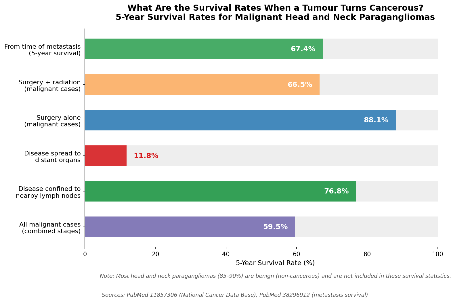 Figure 5: Frequency of genetic mutations among head and neck paraganglioma patients who underwent testing. SDHD is the most commonly identified mutation, followed by SDHB. Data from published genetic screening cohorts.
Because of all this, genetic testing is now strongly recommended for every person diagnosed with a head and neck paraganglioma, regardless of family history or age. If a mutation is found, first-degree relatives (parents, siblings, children) should be offered testing and, if positive, regular screening for tumour development. In SDHD-positive families, the mutation follows a pattern called genomic imprinting: it generally only causes disease when inherited from the father rather than the mother, which affects how family screening is structured.
Clinical predictors that increase the likelihood of finding a germline mutation include: a family history of paraganglioma or adrenal phaeochromocytoma (its adrenal counterpart), multiple tumours at diagnosis, age 40 or younger, and male sex.
What This All Means for Patients
If you have been told you have a head and neck paraganglioma, or are being investigated for one, here are the key points to carry away:
- Most of these tumours are benign. The overwhelming majority do not spread, and with appropriate treatment, the long-term outlook is very good
- Slow growth is the norm. These tumours typically grow about 1 mm per year. There is rarely an emergency, and taking time to plan treatment carefully at a specialist centre is strongly advisable
- Treatment choice depends heavily on location. Carotid body tumours are usually treated surgically; jugular and skull base tumours are increasingly managed with radiation to avoid nerve damage
- Radiation and surgery achieve similar control rates for most tumour types, but radiation preserves quality of life better for skull base tumours
- Genetic testing matters. Even without a family history, a significant proportion of patients carry a hereditary mutation. Knowing this changes follow-up plans and protects family members
- Lifelong surveillance is not optional. Recurrences can and do appear more than a decade after treatment. Regular imaging is essential indefinitely
- Seek a multidisciplinary specialist centre. Because these tumours are rare and complex, outcomes are substantially better in high-volume units with experienced surgeons, radiation oncologists, and skull base specialists working together
Further Reading
Understanding Paragangliomas
- Paraganglioma: StatPearls (NCBI Bookshelf) — comprehensive clinical overview from the NIH database, covering all types and locations
- Paraganglioma: Causes, Symptoms and Treatment (Cleveland Clinic) — accessible patient-facing summary from a leading specialist centre
- Paraganglioma: Symptoms and Causes (Mayo Clinic) — clear lay overview covering what these tumours are and how they present
- Paraganglioma: Diagnosis and Treatment (Mayo Clinic) — companion page covering the investigation and management process
Carotid Body Tumours Specifically
- Carotid Body Tumour: Symptoms, Causes and Treatment (Cleveland Clinic) — dedicated resource focusing on the most common subtype, with information on surgical approaches
Imaging and Diagnosis
- Multimodality Imaging of Paragangliomas of the Head and Neck (PMC) — in-depth review of imaging techniques, including the "salt and pepper" MRI sign and functional imaging
- 68Ga-DOTATATE PET/CT in Head and Neck Paragangliomas (PMC) — explains the role of the most advanced imaging tool now used for whole-body staging
Genetics and Family Screening
- Head and Neck Paragangliomas: Clinical and Molecular Genetic Classification (PubMed) — landmark paper describing the genetic landscape and nine susceptibility genes
- Genetic Testing in Head and Neck Paraganglioma (PubMed) — evidence base for why genetic testing should be offered to all patients
- Natural History and Management of Familial Paraganglioma Syndrome Type 1 (PMC) — focuses on SDHD-linked disease and family management
Treatment Options
- Paragangliomas of the Head and Neck: A Review of Latest Diagnostic and Treatment Methods (PMC) — up-to-date 2024 review covering all treatment modalities and their evidence base
- Active Surveillance Management of Head and Neck Paragangliomas (PubMed) — case series and literature review supporting the watch-and-scan approach
- Efficacy and Safety of Radiotherapy: A Retrospective 23-Year Analysis (PMC) — large, long-term study demonstrating excellent outcomes and low toxicity with modern radiotherapy
Prognosis and Outcomes
- Recurrence and Progression of Head and Neck Paragangliomas After Treatment (PubMed) — data on long-term recurrence rates, supporting the case for lifelong surveillance
- National Cancer Data Base Report on Malignant Paragangliomas of the Head and Neck (PubMed) — large dataset on survival outcomes for the malignant minority
References/Helpful Resources
- Cleveland Clinic. Paraganglioma: Causes, Symptoms and Treatment [Internet]. [cited 2026 Jul 03]. Available from: https://my.clevelandclinic.org/health/diseases/22394-paraganglioma
- Mayo Clinic. Paraganglioma: Symptoms and Causes [Internet]. [cited 2026 Jul 03]. Available from: https://www.mayoclinic.org/diseases-conditions/paraganglioma/symptoms-causes/syc-20575682
- Sandow L, Thawani R, Kim MS, Heinrich MC. Paraganglioma of the Head and Neck: A Review. Endocr Pract. 2023 Feb;29(2):141–7. doi:10.1016/j.eprac.2022.10.002 PubMed PMID: 36252779; PubMed Central PMCID: PMC9979593. Available from: https://pubmed.ncbi.nlm.nih.gov/36252779/
- Crona J, Taïeb D, Pacak K. New Perspectives on Pheochromocytoma and Paraganglioma: Toward a Molecular Classification. Endocr Rev. 2017 Dec 1;38(6):489–515. doi:10.1210/er.2017-00062 PubMed PMID: 28938417; PubMed Central PMCID: PMC5716829. Available from:https://pubmed.ncbi.nlm.nih.gov/28938417/
- Ikram A, Rehman A. Paraganglioma. In: StatPearls [Internet]. Treasure Island (FL): StatPearls Publishing; 2026 [cited 2026 Jul 16]. Available from: http://www.ncbi.nlm.nih.gov/books/NBK549834/ PubMed PMID: 31751024. Available from: https://www.ncbi.nlm.nih.gov/books/NBK549834/
- Offergeld C, Brase C, Yaremchuk S, Mader I, Rischke HC, Gläsker S, et al. Head and neck paragangliomas: clinical and molecular genetic classification. Clinics (Sao Paulo). 2012;67 Suppl 1(Suppl 1):19–28. doi:10.6061/clinics/2012(sup01)05 PubMed PMID: 22584701; PubMed Central PMCID: PMC3328838. Available from: https://pubmed.ncbi.nlm.nih.gov/22584701/
- Palade DO, Hainarosie R, Zamfir A, Vrinceanu D, Pertea M, Tusaliu M, et al. Paragangliomas of the Head and Neck: A Review of the Latest Diagnostic and Treatment Methods. Medicina (Kaunas). 2024 May 30;60(6):914. doi:10.3390/medicina60060914 PubMed PMID: 38929531; PubMed Central PMCID: PMC11205799. Available from: https://pmc.ncbi.nlm.nih.gov/articles/PMC11205799/
- Williams MD. Paragangliomas of the Head and Neck: An Overview from Diagnosis to Genetics. Head Neck Pathol. 2017 Mar 20;11(3):278–87. doi:10.1007/s12105-017-0803-4 PubMed PMID: 28321772; PubMed Central PMCID: PMC5550402. . Available from: https://pmc.ncbi.nlm.nih.gov/articles/PMC5550402/
- Benn DE, Gimenez-Roqueplo AP, Reilly JR, Bertherat J, Burgess J, Byth K, et al. Clinical presentation and penetrance of pheochromocytoma/paraganglioma syndromes. J Clin Endocrinol Metab. 2006 Mar;91(3):827–36. doi:10.1210/jc.2005-1862 PubMed PMID: 16317055. Available from:https://pubmed.ncbi.nlm.nih.gov/16317055/
- Welander J, Söderkvist P, Gimm O. Genetics and clinical characteristics of hereditary pheochromocytomas and paragangliomas. Endocr Relat Cancer. 2011 Dec;18(6):R253-276. doi:10.1530/ERC-11-0170 PubMed PMID: 22041710. Available from: https://pubmed.ncbi.nlm.nih.gov/22041710/
- Cleveland Clinic. Carotid Body Tumour: Symptoms, Causes and Treatment [Internet]. [cited 2026 Jul 03]. Available from: https://my.clevelandclinic.org/health/diseases/16848-carotid-body-tumors
- Urquhart AC, Johnson JT, Myers EN, Schechter GL. Glomus vagale: paraganglioma of the vagus nerve. Laryngoscope. 1994 Apr;104(4):440–5. doi:10.1288/00005537-199404000-00008 PubMed PMID: 8164483. Available from: https://pubmed.ncbi.nlm.nih.gov/8164483/
- Richter S, Pacak K, Kunst HPM, Januszewicz A, Nölting S, Remde H, et al. Management and follow-up strategies for patients with head and neck paraganglioma. Eur J Endocrinol. 2024 Sep 20;191(4):389–98. doi:10.1093/ejendo/lvae113 PubMed PMID: 39303070; PubMed Central PMCID: PMC11443905. .Available from:https://pmc.ncbi.nlm.nih.gov/articles/PMC11443905/
- Mayo Clinic. Paraganglioma: Diagnosis and Treatment [Internet]. [cited 2026 Jul 03]. Available from: https://www.mayoclinic.org/diseases-conditions/paraganglioma/diagnosis-treatment/drc-20575710
- Alduraibi A, Bin Essa N, Alruwaili AA, Bomanji J. Ga-68 DOTATATE PET imaging in the management of head and neck paragangliomas: a potential game changer. Front Oncol. 15:1675974. doi:10.3389/fonc.2025.1675974 PubMed PMID: 41341385; PubMed Central PMCID: PMC12668995. Available from: https://www.ncbi.nlm.nih.gov/pmc/articles/PMC12668995/
- Sridhara SK, Yener M, Hanna EY, Rich T, Jimenez C, Kupferman ME. Genetic testing in head and neck paraganglioma: who, what, and why? J Neurol Surg B Skull Base. 2013 Aug;74(4):236–40. doi:10.1055/s-0033-1342924 PubMed PMID: 24436918; PubMed Central PMCID: PMC3715610. Available from: https://pubmed.ncbi.nlm.nih.gov/24436918/
- Dixon JL, Atkins MD, Bohannon WT, Buckley CJ, Lairmore TC. Surgical management of carotid body tumours: a 15-year single institution experience employing an interdisciplinary approach. Proc (Bayl Univ Med Cent). 2016 Jan;29(1):16–20. doi:10.1080/08998280.2016.11929343 PubMed PMID: 26722157; PubMed Central PMCID: PMC4677842. Available from:https://pmc.ncbi.nlm.nih.gov/articles/PMC4677842/
- Lim JY, Kim J, Kim SH, Lee S, Lim YC, Kim JW, et al. Surgical treatment of carotid body paragangliomas: outcomes and complications according to the Shamblin classification. Clin Exp Otorhinolaryngol. 2010 Jun;3(2):91–5. doi:10.3342/ceo.2010.3.2.91 PubMed PMID: 20607078; PubMed Central PMCID: PMC2896739. Available from: https://pubmed.ncbi.nlm.nih.gov/20607078/
- Suárez C, Rodrigo JP, Bödeker CC, Llorente JL, Silver CE, Jansen JC, et al. Jugular and vagal paragangliomas: Systematic study of management with surgery and radiotherapy. Head Neck. 2013 Aug;35(8):1195–204. doi:10.1002/hed.22976 PubMed PMID: 22422597. Available from:https://pubmed.ncbi.nlm.nih.gov/22422597/
- Ivan ME, Sughrue ME, Clark AJ, Kane AJ, Aranda D, Barani IJ, et al. A meta-analysis of tumour control rates and treatment-related morbidity for patients with glomus jugulare tumours. J Neurosurg. 2011 May;114(5):1299–305. doi:10.3171/2010.9.JNS10699 PubMed PMID: 21029039. Available from: https://pubmed.ncbi.nlm.nih.gov/21029039/
- Jiang X, Fang G, Guo D, Xu X, Chen B, Jiang J, et al. Surgical Management of Carotid Body Tumour and Risk Factors of Postoperative Cranial Nerve Injury. World J Surg. 2020 Dec;44(12):4254–60. doi:10.1007/s00268-020-05723-8 PubMed PMID: 32804251. Available from: https://pubmed.ncbi.nlm.nih.gov/32804251/
- Neskey DM, Hatoum G, Modh R, Civantos F, Telischi FF, Angeli SI, et al. Outcomes after surgical resection of head and neck paragangliomas: a review of 61 patients. Skull Base. 2011 May;21(3):171–6. doi:10.1055/s-0031-1275251 PubMed PMID: 22451821; PubMed Central PMCID: PMC3312103. Available from: https://pubmed.ncbi.nlm.nih.gov/22451821/
- Jackson RS, Myhill JA, Padhya TA, McCaffrey JC, McCaffrey TV, Mhaskar RS. The Effects of Preoperative Embolization on Carotid Body Paraganglioma Surgery: A Systematic Review and Meta-analysis. Otolaryngol Head Neck Surg. 2015 Dec;153(6):943–50. doi:10.1177/0194599815605323 PubMed PMID: 26378186. Available from: https://pubmed.ncbi.nlm.nih.gov/26378186/
- Dupin C, Lang P, Dessard-Diana B, Simon JM, Cuenca X, Mazeron JJ, et al. Treatment of head and neck paragangliomas with external beam radiation therapy. Int J Radiat Oncol Biol Phys. 2014 Jun 1;89(2):353–9. doi:10.1016/j.ijrobp.2014.02.010 PubMed PMID: 24685154. Available from: https://pubmed.ncbi.nlm.nih.gov/24685154/
- Guss ZD, Batra S, Limb CJ, Li G, Sughrue ME, Redmond K, et al. RADIOSURGERY OF GLOMUS JUGULARE TUMORS: A META-ANALYSIS. Int J Radiat Oncol Biol Phys. 2011 Nov 15;81(4):e497–502. doi:10.1016/j.ijrobp.2011.05.006 PubMed PMID: 21703782; PubMed Central PMCID: PMC4086637 Available from:https://pmc.ncbi.nlm.nih.gov/articles/PMC4086637/
- Daza-Ovalle A, Bin-Alamer O, Wei Z, Abou-Al-Shaar H, Hadjipanayis CG, Gardner P, et al. Long-Term Outcomes of Jugular Paragangliomas Undergoing Gamma Knife Radiosurgery: A Single Centre Experience. Neurosurgery. 2025 Jan 29;97(2):463–71. doi:10.1227/neu.0000000000003357 PubMed PMID: 39878475. Available from: https://pubmed.ncbi.nlm.nih.gov/39878475/
- Hinerman RW, Amdur RJ, Morris CG, Kirwan J, Mendenhall WM. Definitive radiotherapy in the management of paragangliomas arising in the head and neck: a 35-year experience. Head Neck. 2008 Nov;30(11):1431–8. doi:10.1002/hed.20885 PubMed PMID: 18704974. Available from: https://pubmed.ncbi.nlm.nih.gov/18704974/
- Jansen JC, van den Berg R, Kuiper A, van der Mey AG, Zwinderman AH, Cornelisse CJ. Estimation of growth rate in patients with head and neck paragangliomas influences the treatment proposal. Cancer. 2000 Jun 15;88(12):2811–6. PubMed PMID: 10870065. Available from:https://pubmed.ncbi.nlm.nih.gov/10870065/
- Langerman A, Athavale SM, Rangarajan SV, Sinard RJ, Netterville JL. Natural history of cervical paragangliomas: outcomes of observation of 43 patients. Arch Otolaryngol Head Neck Surg. 2012 Apr;138(4):341–5. doi:10.1001/archoto.2012.37 PubMed PMID: 22431860. Available from: https://pubmed.ncbi.nlm.nih.gov/22431860/
- Obholzer RJ, Hornigold R, Connor S, Gleeson MJ. Classification and management of cervical paragangliomas. Ann R Coll Surg Engl. 2011 Nov;93(8):596–602. doi:10.1308/147870811X13137608455172 PubMed PMID: 22041235; PubMed Central PMCID: PMC3566684. Available from: https://pubmed.ncbi.nlm.nih.gov/22041235/
- Contrera KJ, Yong V, Reddy CA, Liu SW, Lorenz RR. Recurrence and Progression of Head and Neck Paragangliomas after Treatment. Otolaryngol Head Neck Surg. 2020 Apr;162(4):504–11. doi:10.1177/0194599820902702 PubMed PMID: 32066331. Available from: https://pubmed.ncbi.nlm.nih.gov/32066331/
- Ricketts CJ, Forman JR, Rattenberry E, Bradshaw N, Lalloo F, Izatt L, et al. Tumor risks and genotype-phenotype-proteotype analysis in 358 patients with germline mutations in SDHB and SDHD. Hum Mutat. 2010 Jan;31(1):41–51. doi:10.1002/humu.21136 PubMed PMID: 19802898. Available from: https://pubmed.ncbi.nlm.nih.gov/19802898/
- Schiavi F, Boedeker CC, Bausch B, Peçzkowska M, Gomez CF, Strassburg T, et al. Predictors and prevalence of paraganglioma syndrome associated with mutations of the SDHC gene. JAMA. 2005 Oct 26;294(16):2057–63. doi:10.1001/jama.294.16.2057 PubMed PMID: 16249420. Available from: https://pubmed.ncbi.nlm.nih.gov/16249420/
- Snezhkina AV, Fedorova MS, Pavlov VS, Kalinin DV, Golovyuk AL, Pudova EA, et al. Mutation Frequency in Main Susceptibility Genes Among Patients With Head and Neck Paragangliomas. Front Genet. 2020 Dec 18;11:614908. doi:10.3389/fgene.2020.614908 PubMed PMID: 33391357; PubMed Central PMCID: PMC7775293. Available from: https://www.ncbi.nlm.nih.gov/pmc/articles/PMC7775293/
- Lee JH, Barich F, Karnell LH, Robinson RA, Zhen WK, Gantz BJ, et al. National Cancer Data Base report on malignant paragangliomas of the head and neck. Cancer. 2002 Feb 1;94(3):730–7. doi:10.1002/cncr.10252 PubMed PMID: 11857306.Available from:https://pubmed.ncbi.nlm.nih.gov/11857306/
- Ricketts CJ, Forman JR, Rattenberry E, Bradshaw N, Lalloo F, Izatt L, et al. Tumor risks and genotype-phenotype-proteotype analysis in 358 patients with germline mutations in SDHB and SDHD. Hum Mutat. 2010 Jan;31(1):41–51. doi:10.1002/humu.21136 PubMed PMID: 19802898. Available from: https://pubmed.ncbi.nlm.nih.gov/19802898/
- Contrera KJ, Yong V, Reddy CA, Liu SW, Lorenz RR. Recurrence and Progression of Head and Neck Paragangliomas after Treatment. Otolaryngol Head Neck Surg. 2020 Apr;162(4):504–11. doi:10.1177/0194599820902702 PubMed PMID: 32066331. Available from: https://pubmed.ncbi.nlm.nih.gov/32066331/
