

Most people have never heard of the thymus gland until something goes wrong with it. Tucked behind your breastbone, this small butterfly-shaped organ quietly shapes your immune system during childhood before shrinking into near-irrelevance by adulthood. That gradual retreat, however, doesn't make it immune to disease. Thymoma, a rare tumour that grows from the thymus's lining cells, sits at the intersection of cancer medicine, immunology, and radiology in ways that make it one of the more diagnostically complex conditions a chest specialist will encounter.
If you or someone you know has been told there is a "thymic mass" on a scan, you are likely about to navigate a diagnostic process involving imaging, tissue sampling, and detailed laboratory analysis. This article walks through each of those steps clearly: what each test looks for, why it matters, and what the results actually mean.
What Is Thymoma and Why Does It Matter?
Thymoma is a tumour of the thymus gland's epithelial (lining) cells. It is genuinely rare: roughly 2.2 cases per million people per year in the United States, rising slowly over recent decades, most likely because improved imaging is catching tumours that would previously have gone unnoticed.
Despite its rarity, thymoma punches above its weight diagnostically for two reasons.
First, it frequently presents without any symptoms. Up to 30-50% of thymomas are discovered incidentally on a chest X-ray or CT scan ordered for something entirely unrelated. When symptoms do appear, they typically involve chest pressure, cough, or shortness of breath caused by the tumour pressing on nearby structures.
Second, thymoma has an unusual relationship with the immune system. Because the thymus is where T-cells (immune cells) mature, a cancerous thymus can produce abnormal T-cells that turn on the body itself. This leads to autoimmune complications, the most common being myasthenia gravis, a condition causing progressive muscle weakness. Around 30-50% of thymoma patients also develop myasthenia gravis, and conversely, if you have myasthenia gravis, there is approximately a 1-in-5 chance you have a thymoma.
This bidirectional relationship means two quite different groups of people end up being investigated for thymoma: those with an unexpected chest finding, and those whose neurologist notices something worrying in a muscle weakness workup.
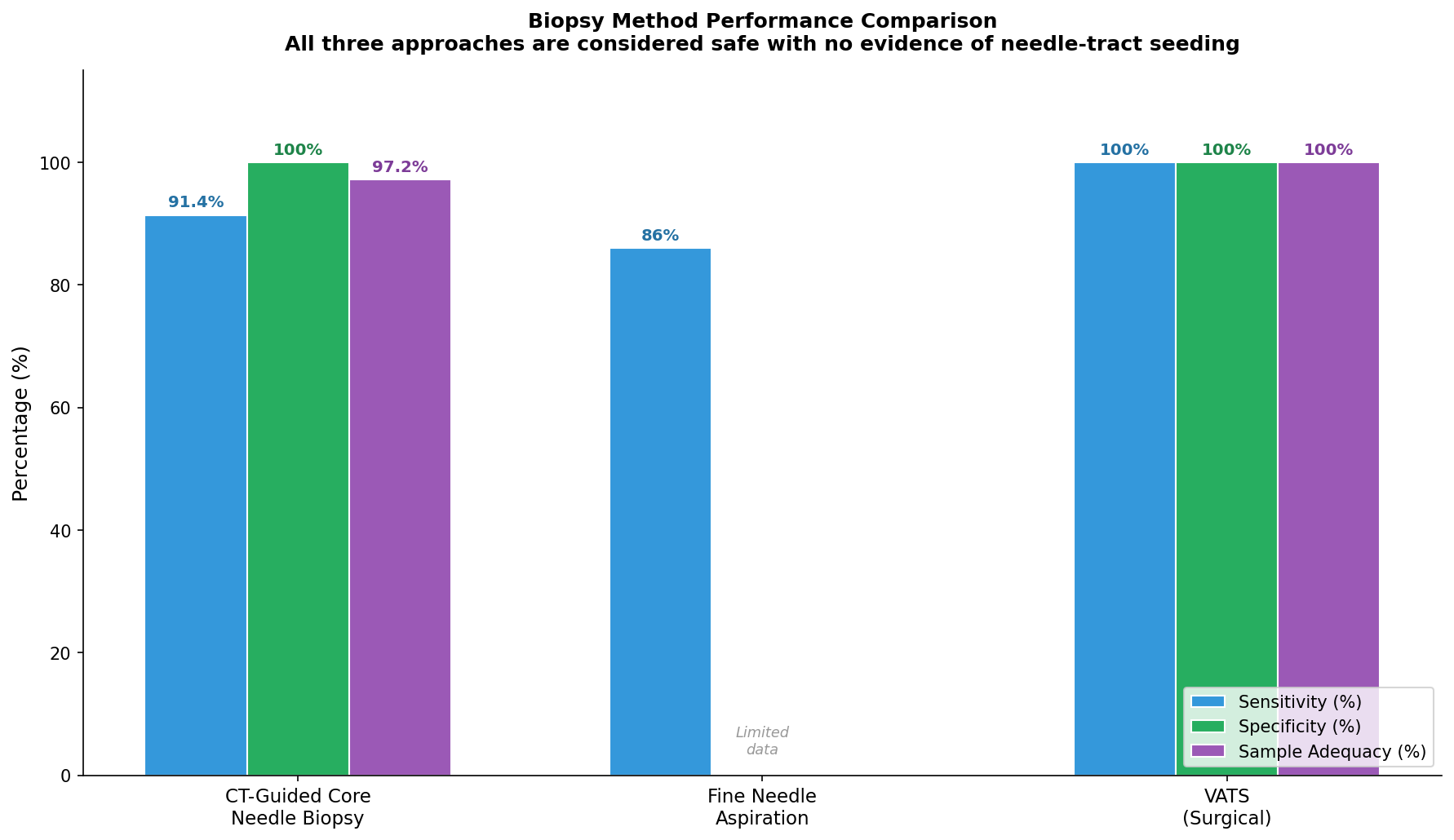 Figure 1: The range of autoimmune conditions (paraneoplastic syndromes) that can occur alongside thymoma. Myasthenia gravis accounts for the majority of cases. Data from a systematic review published on PubMed (PMID 31982129).
The Diagnostic Journey: An Overview
Diagnosing thymoma is rarely a single event. It unfolds in overlapping phases:
- Clinical assessment: History, symptoms, and any autoimmune features
- Imaging: CT scan first, then MRI or PET/CT if additional information is needed
- Tissue sampling (biopsy): To confirm the diagnosis and classify the tumour type
- Histopathological evaluation: Microscopic and molecular analysis of tumour cells
- Staging: Combining imaging and pathology to determine how far the tumour has spread
Each phase answers specific questions. Imaging tells you where the tumour is and what it looks like. A biopsy tells you what it is made of. Histopathology classifies which type it is and how aggressive it is likely to be. Staging combines all of this into a treatment roadmap.
What Imaging Shows: CT, MRI, and PET/CT
CT Scanning: The First-Line Investigation
A contrast-enhanced CT scan of the chest is the standard first-line imaging tool for any suspected thymic mass. It is fast, widely available, and extraordinarily informative about a tumour's size, shape, and relationship to the heart, major blood vessels, and lungs.
On CT, a typical early-stage thymoma appears as a smooth, oval, well-defined mass sitting in the anterior mediastinum (the front compartment of the chest cavity, behind the breastbone). It enhances evenly when contrast dye is injected, meaning it lights up uniformly. About one-third of thymomas show heterogeneous (uneven) enhancement due to areas of bleeding or cysts within the tumour. Some thymomas have thin, peripheral calcification visible at their edges.
What the CT is really looking for are signs of invasion: does the tumour respect its capsule, or does it push into surrounding fat, the heart lining (pericardium), the great vessels, or the lung? These features directly determine the tumour's Masaoka-Koga stage (explained below) and whether surgery will be straightforward or complex.
The diagnostic accuracy of CT for detecting thymoma is high: sensitivity ranges from 85-97% depending on the clinical context, with specificity similarly in the 75-98.7% range.
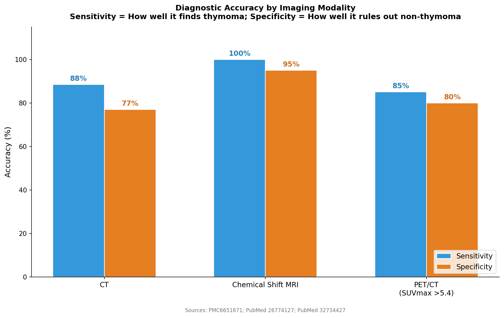 Figure 2: Comparison of sensitivity, specificity, and clinical role across CT, MRI, and PET/CT imaging modalities for thymoma. Sources: PubMed PMID 16905385, PMC6651671, PubMed PMID 26545017.
MRI: When You Need a Clearer Picture
MRI is used as a second-line or complementary tool, particularly when CT findings are ambiguous or when the patient cannot receive CT contrast dye (due to kidney problems or allergy).
Standard MRI sequences are useful, but where MRI genuinely outperforms CT is in a specialist technique called chemical shift MRI (CS-MRI). This exploits the difference in magnetic behaviour between tumour tissue and fat-containing tissue. A particular ratio called the Chemical Shift Ratio (CSR) achieves 100% sensitivity and 95% specificity for distinguishing thymoma from normal or hyperplastic (enlarged but benign) thymus tissue. In comparison, CT achieves only 90.9% sensitivity and 70% specificity for the same task. Crucially, chemical shift MRI does not require contrast injection.
For patients with myasthenia gravis being investigated for an underlying thymoma, MRI overall accuracy is significantly better than CT: 94.3% versus 83.0%.
Diffusion-weighted MRI (DWI) is another advanced technique that measures how freely water molecules move through tissue. In denser, more cellular tumours (typically more aggressive), water diffusion is restricted and generates a measurable signal. Studies show DWI can help distinguish low-risk from high-risk thymoma (AUROC 0.804) and thymoma from thymic carcinoma (AUROC 0.851).
PET/CT: Measuring Metabolic Activity
PET/CT uses a radioactive glucose tracer (18F-FDG) to measure how metabolically active a tumour is. More aggressive, rapidly dividing tumours consume more glucose and therefore "light up" more brightly. This brightness is measured as SUVmax (standardised uptake value maximum).
The pattern is clinically meaningful:
- Thymic hyperplasia (benign enlargement): SUVmax approximately 1.1
- Low-risk thymoma: SUVmax approximately 3.0
- High-risk thymoma: SUVmax approximately 4.42-6.01
- Thymic carcinoma (the most aggressive thymic tumour): SUVmax approximately 7.0-9.09
An SUVmax cut-off of 5.40 achieves 85% sensitivity and 80% specificity for identifying advanced stage (Masaoka-Koga III/IV) disease. A cut-off of 5.60 achieves 89% sensitivity and 78% specificity for WHO malignancy grading.
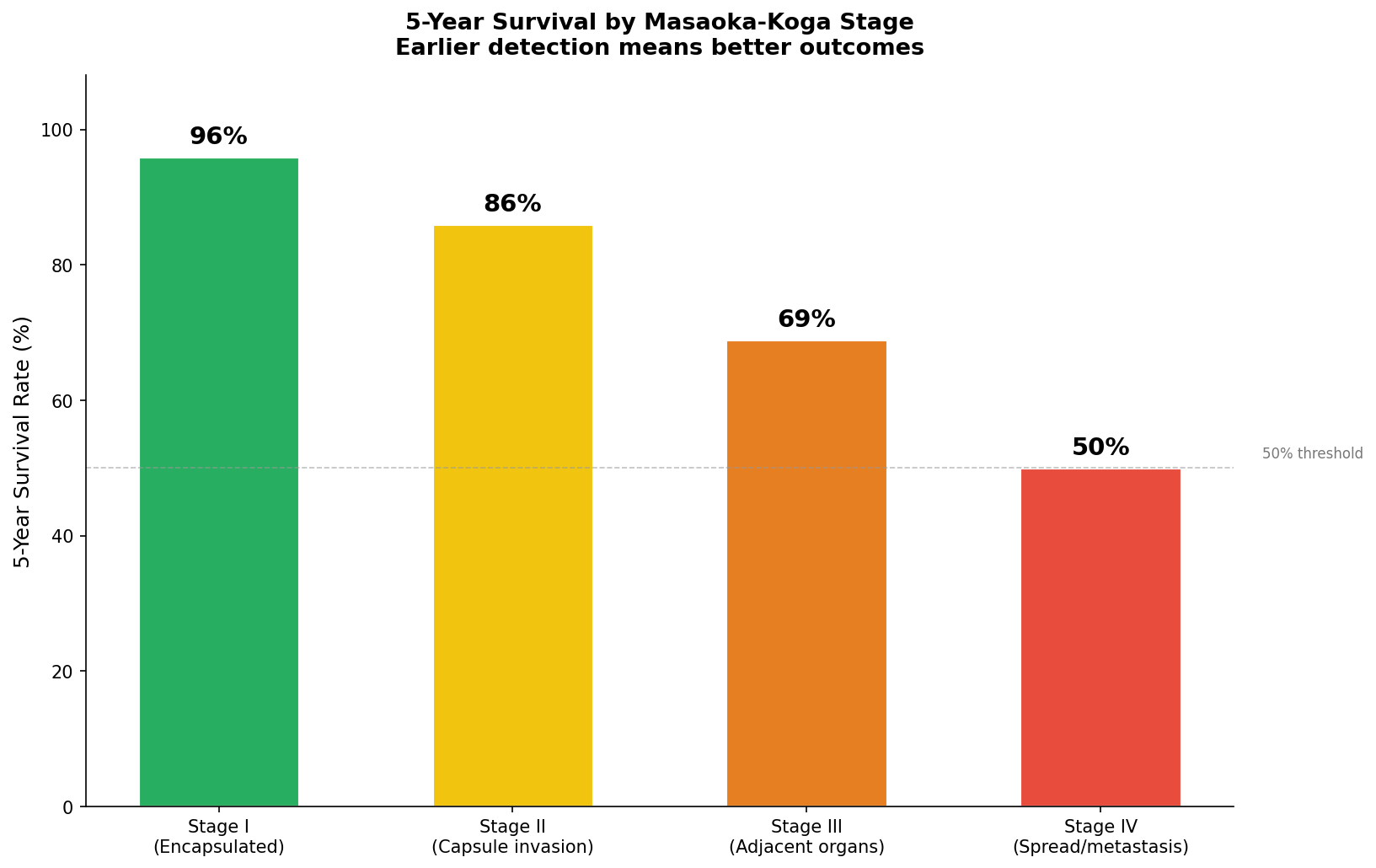 Figure 3: Mean SUVmax values from FDG PET/CT scanning across different thymic tumour subtypes, from thymic hyperplasia through to thymic carcinoma. Higher values indicate a more metabolically active, aggressive disease. Sources: PubMed PMID 21403583, PMID 32734427, PMC5221681.
PET/CT is particularly useful for staging (identifying distant spread) and for monitoring response to treatment. Its limitation is that it cannot reliably distinguish invasive from non-invasive thymoma in the lower SUVmax range: the evidence remains equivocal on this specific question.
Getting a Tissue Sample: How Biopsy Works
No imaging test, however sophisticated, can substitute for looking at actual tumour cells under a microscope. Biopsy is the step that confirms the diagnosis and, critically, determines which type of thymoma it is, information that directly guides treatment decisions.
CT-Guided Core Needle Biopsy: The Preferred Approach
The most commonly used technique is a CT-guided percutaneous core needle biopsy. Under CT imaging guidance, a radiologist inserts a thin needle through the chest wall into the tumour and removes small cylinders of tissue (cores). The procedure takes around 30-60 minutes under local anaesthetic and does not require general anaesthesia or an overnight hospital stay.
The diagnostic performance of this technique is strong:
- Sensitivity for thymic tumours: 89.4-93.3%
- Specificity: 100%
- Overall accuracy: 93.5%
- Sample adequacy (enough tissue for analysis): 97.2% of biopsies
- Concordance with final surgical specimen for WHO subtype classification: 79.4%
Immediate complications (mainly minor bleeding or pneumothorax, a small air leak) occur in approximately 4.4% of cases, typically resolving without intervention.
What About the Risk of Seeding?
A longstanding concern with biopsying thymoma is whether the needle might inadvertently scatter cancer cells along its tract through the chest wall ("seeding"). This is a legitimate question, and the evidence is reassuring. A study with a median follow-up of nearly 39 months found no needle tract seeding in patients who underwent CT-guided core biopsy. One historical case report documented chest wall seeding 12 years after biopsy, but modern analyses attribute pleural seeding in thymoma to tumour biology and incomplete surgical resection, not to the biopsy needle itself.
The consensus from current evidence is that core needle biopsy is accurate and safe for thymoma, and should be offered when tissue diagnosis is required before treatment.
Fine Needle Aspiration: Less Tissue, Less Information
Fine needle aspiration (FNA) uses a finer needle to extract individual cells rather than tissue cores. It is quicker and carries a marginally lower risk, but provides a much smaller cellular sample without preserving the tumour's architectural structure. FNA diagnosis concurs with histological confirmation in approximately 86% of cases, but it cannot reliably determine the WHO histological subtype because subtyping depends on seeing how the cells are arranged relative to each other, not just what individual cells look like.
FNA remains useful for confirming malignancy and ruling out other diagnoses (such as lymphoma) when a core biopsy is not possible.
Surgical Biopsy (VATS): The High-Yield Option
Video-assisted thoracoscopic surgery (VATS) is a minimally invasive surgical approach where a camera and instruments are inserted through small incisions in the chest wall. Used as a biopsy method, it achieves a 100% diagnostic yield: in one surgical series, histological diagnosis was confirmed in every case. It also allows the surgeon to directly visualise the tumour and take larger tissue samples.
VATS biopsy is typically reserved for cases where needle biopsy has been inconclusive or where the surgeon plans to proceed directly to resection once the diagnosis is confirmed intraoperatively.
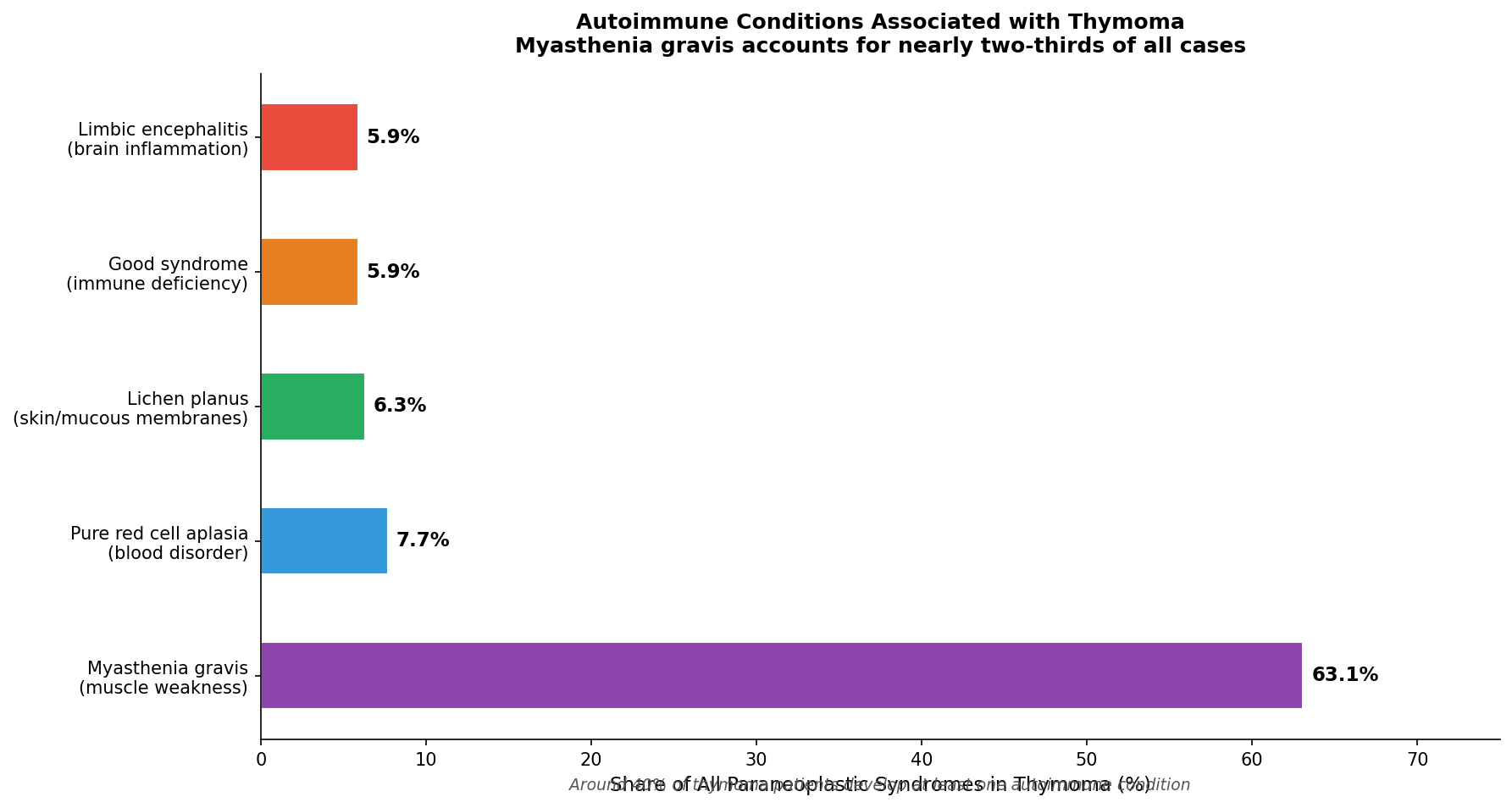 Figure 4: Comparison of diagnostic accuracy, invasiveness, and key features of the three main biopsy approaches for thymoma: CT-guided core needle biopsy, fine needle aspiration, and VATS. Sources: PubMed PMID 16905385, PMID 26397325, PMID 38220515, PMID 28872098.
What the Lab Sees: Histopathological Evaluation
Once tissue is obtained, a pathologist examines it under a microscope and applies a battery of laboratory tests. This is where the diagnosis truly crystallises into something actionable.
The WHO Classification System: Why Tumour Type Predicts Outcome
The World Health Organisation classifies thymomas into five main subtypes (A, AB, B1, B2, B3) based on two key microscopic features: what the epithelial (lining) cells look like, and how many lymphocytes (immune cells) surround them. The classification runs from least to most aggressive.
Type A thymoma has spindle-shaped epithelial cells with bland, innocuous-looking nuclei and very few surrounding lymphocytes. It is the most benign form, with a 100% 20-year survival and an invasiveness rate of only 23.8%.
Type AB thymoma has a mixed appearance, combining spindle-shaped regions with lymphocyte-rich zones. Twenty-year survival is 87%, and just over half (51%) show some invasive behaviour.
Type B1 thymoma closely resembles normal thymic tissue. The epithelial cells are plump and rounded, surrounded by abundant immature T-lymphocytes (called TdT-positive lymphocytes under IHC staining). Despite a high invasiveness rate (73.3%), 20-year survival remains good at 91%.
Type B2 thymoma has more prominent epithelial cells with mild nuclear irregularity, fewer lymphocytes, and characteristic perivascular spaces. Twenty-year survival drops to 65%, and 82% show invasive behaviour.
Type B3 thymoma is the most aggressive thymoma proper. The epithelial cells are polygonal with moderate nuclear atypia, arranged in sheets. All B3 thymomas (100%) behave invasively, and 20-year survival falls to only 38%.
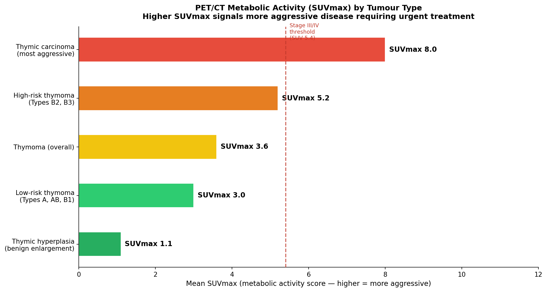 Figure 5: Twenty-year survival rates by WHO histological subtype, from the most indolent (Type A, 100%) to the most aggressive within the thymoma category (Type B3, 38%). Data from PubMed PMID 12048910, PMID 11145244.
The Masaoka-Koga Staging System: How Far Has It Spread?
Alongside the WHO classification, every thymoma is assigned a Masaoka-Koga stage that describes how much the tumour has spread beyond its capsule:
- Stage I: Fully encapsulated with no invasion. Five-year survival: 96%
- Stage II: Microscopic invasion through the capsule into the surrounding fat or adhesion to the pleura. Five-year survival: 86%
- Stage III: Visible invasion into adjacent organs (pericardium, major blood vessels, lung). Five-year survival: 69%
- Stage IV: Spread to the pleura, pericardium (IVa), or through lymph/blood vessels to distant sites (IVb). Five-year survival: 50%
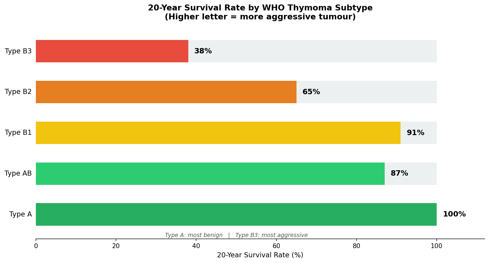 Figure 6: Five-year and 10-year overall survival rates by Masaoka-Koga stage. The stepwise decline underscores why identifying invasion accurately on imaging is so clinically critical. Sources: PMC8582470, PubMed PMID 25534526.
Crucially, complete surgical removal is the single most important prognostic factor for thymoma. Patients with clear surgical margins have a 5-year survival of 86% compared to 28% for those where complete removal was not achieved.
Immunohistochemistry: The Molecular Fingerprint
Simply looking at cell shape under a microscope is not always enough. Immunohistochemistry (IHC) applies specific antibodies to tumour tissue sections that bind to particular proteins, revealing which proteins the cells express and don't express. This helps distinguish thymoma subtypes from each other and from entirely different diagnoses (like lymphoma or thymic carcinoma) that might look similar.
Key IHC markers include:
- CD5: Negative in thymoma; positive in approximately 70% of thymic carcinomas. This is a critical distinguishing feature
- CD117 (also called KIT): Positive in only 4% of thymomas but 80% of thymic carcinomas
- TdT: Marks immature T-lymphocytes. Abundant in B1, moderate in B2, and absent in A and B3
- Beta-5t: The most useful single positive marker for confirming thymoma identity
- Cytokeratin: Positive in all thymoma subtypes, confirming epithelial origin
A four-marker IHC panel (CD117, BAP1, MTAP, TdT) correctly predicts thymic carcinoma in 88.9% of cases and thymoma in 77.8% of cases, even from limited biopsy material. This is particularly valuable when only a small core biopsy sample is available.
Pulling It All Together: How the Diagnosis Is Made
A complete thymoma diagnosis is not a single test result. It is a synthesis:
The CT scan identifies a mass, characterises its borders, looks for signs of invasion, and provides a provisional stage. Chemical shift MRI or PET/CT may add metabolic or tissue-composition data. The biopsy provides actual tumour tissue. The pathologist uses H&E (haematoxylin and eosin) staining to classify the WHO subtype, and IHC to confirm the diagnosis and exclude look-alikes. The Masaoka-Koga stage is then determined by combining imaging findings with surgical or pathological confirmation.
Together, the WHO classification and Masaoka-Koga stage provide a multi-dimensional picture that guides every subsequent decision: whether to operate directly, whether to give chemotherapy first to shrink the tumour, what follow-up imaging schedule to use, and what surveillance is needed for associated autoimmune conditions.
The good news is that the diagnostic toolkit for thymoma is excellent. Even small biopsy specimens can yield reliable diagnoses. Imaging is sufficiently accurate to plan surgery in most cases. And for the many patients whose thymoma is caught at Stage I or II, the long-term outlook after complete surgical removal is genuinely excellent: five-year survival exceeds 95%.
For patients at the start of this diagnostic journey, the process can feel daunting. But each step is purposeful. Every scan, every biopsy, every stain on a glass slide is answering a specific question that brings treatment one step closer.
Further Reading
Understanding Thymoma and the Thymus
- Thymoma and Thymic Carcinoma: Treatment Information (NCI)
- Thymoma and Thymic Carcinoma Overview (Cleveland Clinic)
- MedlinePlus: Thymoma
Imaging of Thymic Tumours
- Imaging Evaluation of Thymoma and Thymic Carcinoma (PMC8762255)
- Update in Diagnostic Imaging of Thymus and Anterior Mediastinal Masses (PMC6755948)
- State of the Art: MRI of Thymoma (PubMed 25952513)
Biopsy and Tissue Diagnosis
- CT-Guided Core Needle Biopsy for Thymic Tumours: Accuracy and Seeding Risk (PubMed 38220515)
- Diagnostic Yield of Percutaneous Needle Biopsy for Anterior Mediastinal Masses (PubMed 26397325)
Histopathology and Classification
- ITMIG Consensus on WHO Classification of Thymoma (PubMed 24722150)
- Diagnostic Roles of Immunohistochemistry in Thymic Tumours (PMC7399973)
Myasthenia Gravis and Paraneoplastic Syndromes
- Myasthenia Gravis and Thymic Neoplasms: A Review (PMC4677085)
- Systematic Review of Paraneoplastic Syndromes in Thymoma (PubMed 31982129)
Staging and Prognosis
- Masaoka-Koga and TNM Staging: Prognostic Comparison (PMC8582470)
- Long-Term Outcomes After Complete Thymoma Resection (PMC5433013)
References/Helpful Resources
- Bushan K, Sharma S, Verma H. A Review of Thymic Tumours. Indian J Surg Oncol. 2013 Jun;4(2):112–6. doi:10.1007/s13193-013-0214-2 PubMed PMID: 24426710; PubMed Central PMCID: PMC3693138.
- National Cancer Institute. Thymoma and Thymic Carcinoma Treatment (PDQ) [Internet]. Bethesda: NCI; [cited 2026 Jul 2]. Available from: https://www.cancer.gov/types/thymoma/patient/thymoma-treatment-pdq
- Cleveland Clinic. Thymoma (Thymic Carcinoma): Symptoms, Causes and Treatment [Internet]. Cleveland: Cleveland Clinic; [cited 2026 Jul 2]. Available from: https://my.clevelandclinic.org/health/diseases/6196-thymoma-and-thymic-carcinoma
- Tuan PA, Vien MV, Dong HV, Sibell D, Giang BV. The Value of CT and MRI for Determining Thymoma in Patients With Myasthenia Gravis. Cancer Control. 2019 Jul 23;26(1):1073274819865281. doi:10.1177/1073274819865281 PubMed PMID: 31331197; PubMed Central PMCID: PMC6651671. Available from: https://pmc.ncbi.nlm.nih.gov/articles/PMC6651671/
- Ettinger DS, Riely GJ, Akerley W, Borghaei H, Chang AC, Cheney RT, et al. Thymomas and thymic carcinomas: Clinical Practice Guidelines in Oncology. J Natl Compr Canc Netw. 2013 May 1;11(5):562–76. doi:10.6004/jnccn.2013.0072 PubMed PMID: 23667206.
- Strange CD, Ahuja J, Shroff GS, Truong MT, Marom EM. Imaging Evaluation of Thymoma and Thymic Carcinoma. Front Oncol. 2022 Jan 3;11:810419. doi:10.3389/fonc.2021.810419 PubMed PMID: 35047412; PubMed Central PMCID: PMC8762255. Available from: https://pmc.ncbi.nlm.nih.gov/articles/PMC8762255/
- Carter BW, Benveniste MFK, Truong MT, Marom EM. State of the Art: MR Imaging of Thymoma. Magn Reson Imaging Clin N Am. 2015 May;23(2):165–77. doi:10.1016/j.mric.2015.01.005 PubMed PMID: 25952513. Available from: https://pubmed.ncbi.nlm.nih.gov/25952513/
- Liu Y. Characterisation of thymic lesions with F-18 FDG PET-CT: an emphasis on epithelial tumours. Nucl Med Commun. 2011 Jul;32(7):554–62. doi:10.1097/MNM.0b013e328345b984 PubMed PMID: 21403583. Available from: https://pubmed.ncbi.nlm.nih.gov/21403583/
- Park SY, Cho A, Bae MK, Lee CY, Kim DJ, Chung KY. Value of 18F-FDG PET/CT for Predicting the World Health Organisation Malignant Grade of Thymic Epithelial Tumours: Focused on Volume-Dependent Parameters. Clin Nucl Med. 2016 Jan;41(1):15–20. doi:10.1097/RLU.0000000000001032 PubMed PMID: 26545017. Available from: https://pubmed.ncbi.nlm.nih.gov/26545017/
- Petranovic M, Gilman MD, Muniappan A, Hasserjian RP, Digumarthy SR, Muse VV, et al. Diagnostic Yield of CT-Guided Percutaneous Transthoracic Needle Biopsy for Diagnosis of Anterior Mediastinal Masses. AJR Am J Roentgenol. 2015 Oct;205(4):774–9. doi:10.2214/AJR.15.14442 PubMed PMID: 26397325. Available from: https://pubmed.ncbi.nlm.nih.gov/26397325/
- Ahn Y, Lee SM, Choi S, Choe J, Oh SY, Do KH, et al. CT-guided pretreatment biopsy diagnosis in patients with thymic epithelial tumours: diagnostic accuracy and risk of seeding. Clin Radiol. 2024 Apr;79(4):263–71. doi:10.1016/j.crad.2023.12.009 PubMed PMID: 38220515. Available from: https://pubmed.ncbi.nlm.nih.gov/38220515/
- Okumura M, Ohta M, Miyoshi S, Mori T, Yasumitsu T, Nakahara K, et al. Oncological significance of WHO histological thymoma classification. A clinical study based on 286 patients. Jpn J Thorac Cardiovasc Surg. 2002 May;50(5):189–94. doi:10.1007/BF03032284 PubMed PMID: 12048910. Available from: https://pubmed.ncbi.nlm.nih.gov/12048910/
- Kondo K, Yoshizawa K, Tsuyuguchi M, Kimura S, Sumitomo M, Morita J, et al. WHO histologic classification is a prognostic indicator in thymoma. Ann Thorac Surg. 2004 Apr;77(4):1183–8. doi:10.1016/j.athoracsur.2003.07.042 PubMed PMID: 15063231. Available from: https://pubmed.ncbi.nlm.nih.gov/15063231/
- Nakagawa K, Asamura H, Matsuno Y, Suzuki K, Kondo H, Maeshima A, et al. Thymoma: a clinicopathologic study based on the new World Health Organisation classification. J Thorac Cardiovasc Surg. 2003 Oct;126(4):1134–40. doi:10.1016/s0022-5223(03)00798-0 PubMed PMID: 14566259.
- Chalabreysse L, Roy P, Cordier JF, Loire R, Gamondes JP, Thivolet-Bejui F. Correlation of the WHO schema for the classification of thymic epithelial neoplasms with prognosis: a retrospective study of 90 tumours. Am J Surg Pathol. 2002 Dec;26(12):1605–11. doi:10.1097/00000478-200212000-00008 PubMed PMID: 12459627.
- Marx A, Ströbel P, Badve SS, Chalabreysse L, Chan JKC, Chen G, et al. ITMIG consensus statement on the use of the WHO histological classification of thymoma and thymic carcinoma: refined definitions, histological criteria, and reporting. J Thorac Oncol. 2014 May;9(5):596–611. doi:10.1097/JTO.0000000000000154 PubMed PMID: 24722150. Available from: https://pubmed.ncbi.nlm.nih.gov/24722150/
- Moon JW, Lee KS, Shin MH, Kim S, Woo SY, Lee G, et al. Thymic epithelial tumours: prognostic determinants among clinical, histopathologic, and computed tomography findings. Ann Thorac Surg. 2015 Feb;99(2):462–70. doi:10.1016/j.athoracsur.2014.09.050 PubMed PMID: 25534526. Available from: https://pubmed.ncbi.nlm.nih.gov/2553452/
- Yuan ZY, Gao SG, Mu JW, Xue Q, Mao YS, Wang DL, et al. Long-term outcomes of 307 patients after complete thymoma resection. Chin J Cancer. 2017 May 15;36:46. doi:10.1186/s40880-017-0213-8 PubMed PMID: 28506287; PubMed Central PMCID: PMC5433013. Available from: https://pmc.ncbi.nlm.nih.gov/articles/PMC5433013/
- Dorfman DM, Shahsafaei A, Chan JK. Thymic carcinomas, but not thymomas and carcinomas of other sites, show CD5 immunoreactivity. Am J Surg Pathol. 1997 Aug;21(8):936–40. doi:10.1097/00000478-199708000-00008 PubMed PMID: 9255257. Available from: https://pubmed.ncbi.nlm.nih.gov/9255257/
- Bhora FY, Chen DJ, Detterbeck FC, Asamura H, Falkson C, Filosso PL, et al. The ITMIG/IASLC Thymic Epithelial Tumors Staging Project: A Proposed Lymph Node Map for Thymic Epithelial Tumors in the Forthcoming 8th Edition of the TNM Classification of Malignant Tumors. J Thorac Oncol. 2014 Sep;9(9 Suppl 2):S88-96. doi:10.1097/JTO.0000000000000293 PubMed PMID: 25396317.
- TSUCHIDA M. Efficacy and safety of extended thymectomy for elderly patients with myasthenia gravis. Ann Thorac Surg [Internet]. 1999 [cited 2026 Jul 15];67:1563–7. Available from: https://cir.nii.ac.jp/crid/1570854174295660288?lang=en
