

The ABCDE rule for spotting suspicious moles sounds simple enough: check for asymmetry, irregular borders, colour variation, diameter over 6mm and any changes. Here is what most people do not realise; this decades-old framework is not a diagnostic test, it is a practical early-warning system that has saved hundreds of thousands of lives by catching melanomas early, before it spreads.
This guide explains what the ABCDE criteria is, how it works, where it falls short and most importantly, what you should do when you spot something concerning and why early detection remains the single best option for skin cancer survival.
A Brief History of How We Learned to Spot Melanoma
Before 1985, there was no simple framework for identifying melanoma. Doctors relied on intuition and experience and millions of people had their skin cancer discovered too late. Then, dermatologists created the ABCD rule: four straightforward criteria that non-medical professionals could use to detect symptoms.
Over the decades, this framework has spread globally through public health campaigns, medical school curricula and patient education programs. When researchers added the "E" for Evolution (changes over time), they completed a simple yet surprisingly effective tool for early detection. Today, ABCDE remains the gold standard taught to patients, primary care doctors and even specialist dermatologists.
While it may not catch every melanoma, it fundamentally changed how societies approach skin cancer prevention. Localised melanomas caught through ABCDE screening have a 99.4% five-year survival rate, compared to just 35.6% for melanomas that have already spread. That difference of 63.8% points illustrates why catching the disease early transforms outcomes.
The Scale of the Problem: Why Skin Cancer Screening Matters
Skin cancer is the most common cancer diagnosis in the United States, affecting millions annually. In 2021, melanoma incidence reached 23.0 per 100,000 people, a 52% increase over the two decades since 1999. Globally, around 330,000 people are diagnosed with melanoma annually, with approximately 60,000 deaths.
However, whilst melanoma incidence is rising, melanoma mortality has declined by 26% since 1999, dropping from 2.7 to 2.0 per 100,000 and this is due to better detection through screening programs like ABCDE, combined with effective treatments and increased awareness among at-risk populations.
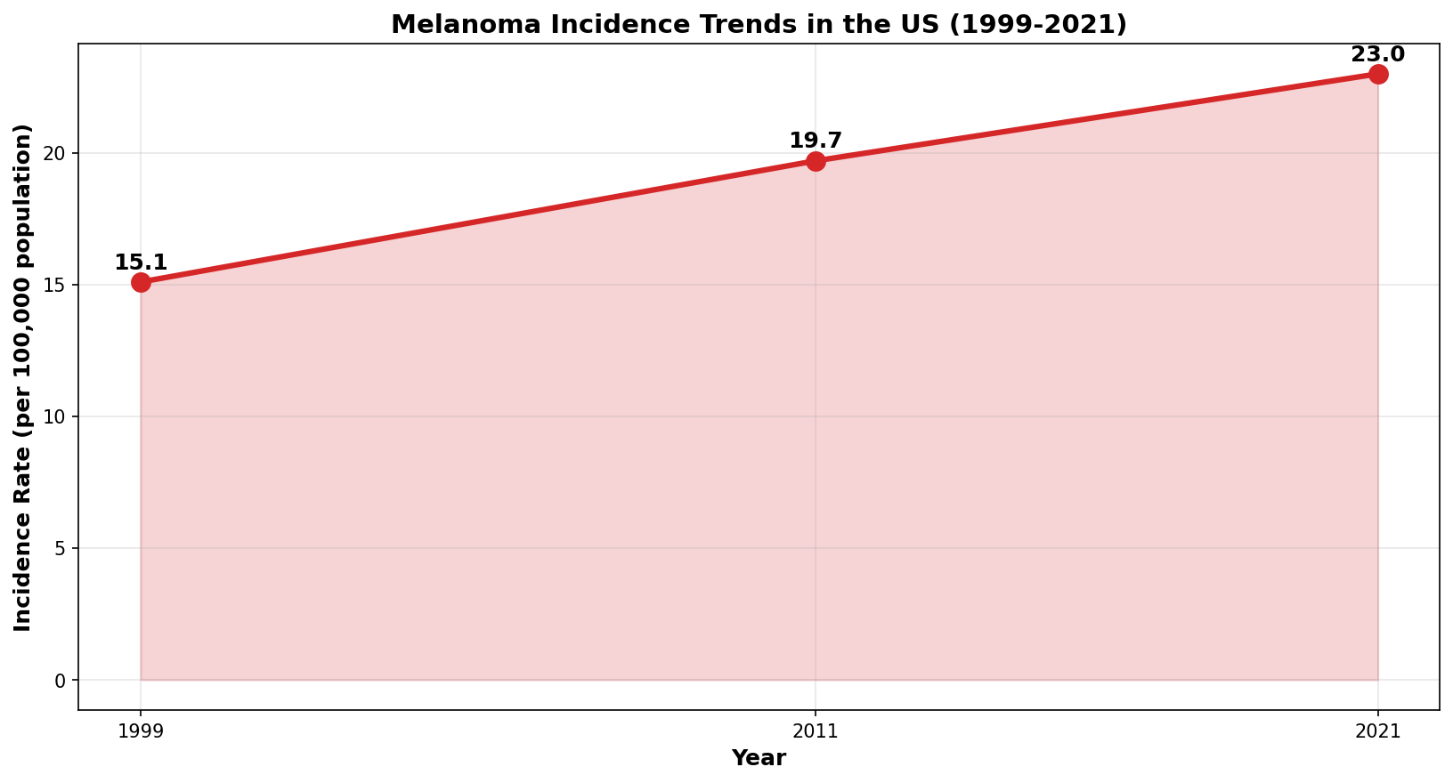
Figure 1: Melanoma incidence in the United States has risen 52% since 1999 (from 15.1 to 23.0 per 100,000), though mortality has declined 26%, indicating earlier detection is working.
Non-melanoma skin cancers (basal cell and squamous cell carcinoma) are even more common, with over 5 million new cases annually in the US alone. These account for more cancer diagnoses than all other cancers combined.
Understanding ABCDE
A for Asymmetry
Asymmetry means one half of the mole does not look like the other half. Imagine drawing an invisible line down the middle of the mole. In a benign mole, both halves would mirror each other fairly evenly, however, in melanoma, they are asymmetrical. Asymmetry is present in only about half of small melanomas (those under 6mm). This means a perfectly symmetrical mole is not necessarily safe. Conversely, an asymmetrical mole could be benign. Asymmetry is one warning sign amongst many and not a definitive answer on its own.
B for Border Irregularity
Borders of benign moles are typically clean, smooth and well-defined. Melanomas are 8.9 times more likely to have irregular borders compared to benign lesions. These might appear jagged, scalloped, notched or blurred, as though the edges fade gradually into surrounding skin rather than stopping abruptly. However, about 54.5% of small melanomas actually have regular borders. Again, the presence or absence of this single feature is not definitive.
C for Colour Variation
Most benign moles are a uniform brown or tan shade throughout. Melanomas often contain multiple colours: browns, tans, blacks, reds, whites or even blues scattered throughout. Yet the statistics reveal nuance: colour variation alone appears in only 63% of melanomas. Three out of ten melanomas are a single colour. However, when researchers look at the uneven distribution of colour (combining colour variation with surface texture changes), the figure jumps to 94% of melanomas, so not just for multiple colours, but also uneven or irregular distribution of colours are present.
D for Diameter
Moles larger than 6mm warrant closer attention. Yet approximately 28% of all melanomas are smaller than 6mm. This is one of the most important limitations of the ABCDE rule. If you assume any small mole is safe, you will miss about one-third of early melanomas. Size alone cannot distinguish benign from malignant, it is just one piece of the puzzle.
E for Evolution
Evolution or any change in the mole over weeks or months might be the most important criterion for patients to monitor. Changes might include growth, shifts in shape, alterations in colour, increase in height or texture or new symptoms like itching, bleeding or tenderness. Benign moles typically remain stable over years. Melanomas, by contrast, grow irregularly and change visibly over short timeframes.
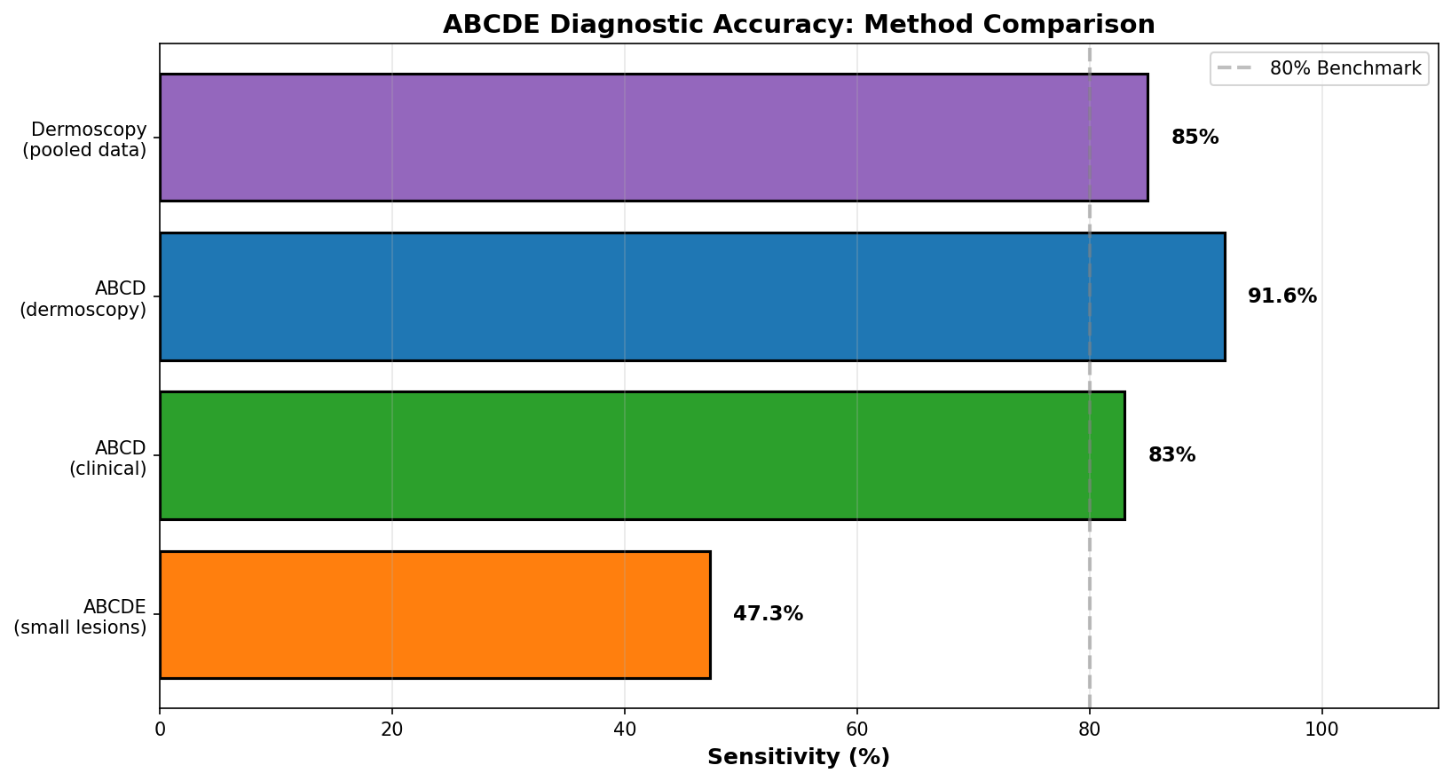
Figure 2: Diagnostic accuracy varies substantially by method. The ABCDE clinical examination achieves 47–83% sensitivity depending on lesion size and examiner expertise, while dermoscopy (magnified examination) improves sensitivity to 84–91.6%.
What Dermatologists Actually Look For
When you present a concerning mole to a dermatologist, they do not simply check off ABCDE criteria because not all melanomas present with all the signs. Asymmetry appears in only 46.3% of small melanomas, borders may be regular in 54.5% of small melanomas, a single colour is present in 60.2% of small melanomas and the diameter over 6mm misses 28% of melanomas entirely. Instead, they consider these features in combination and look at the overall pattern, rather than checking individual boxes.
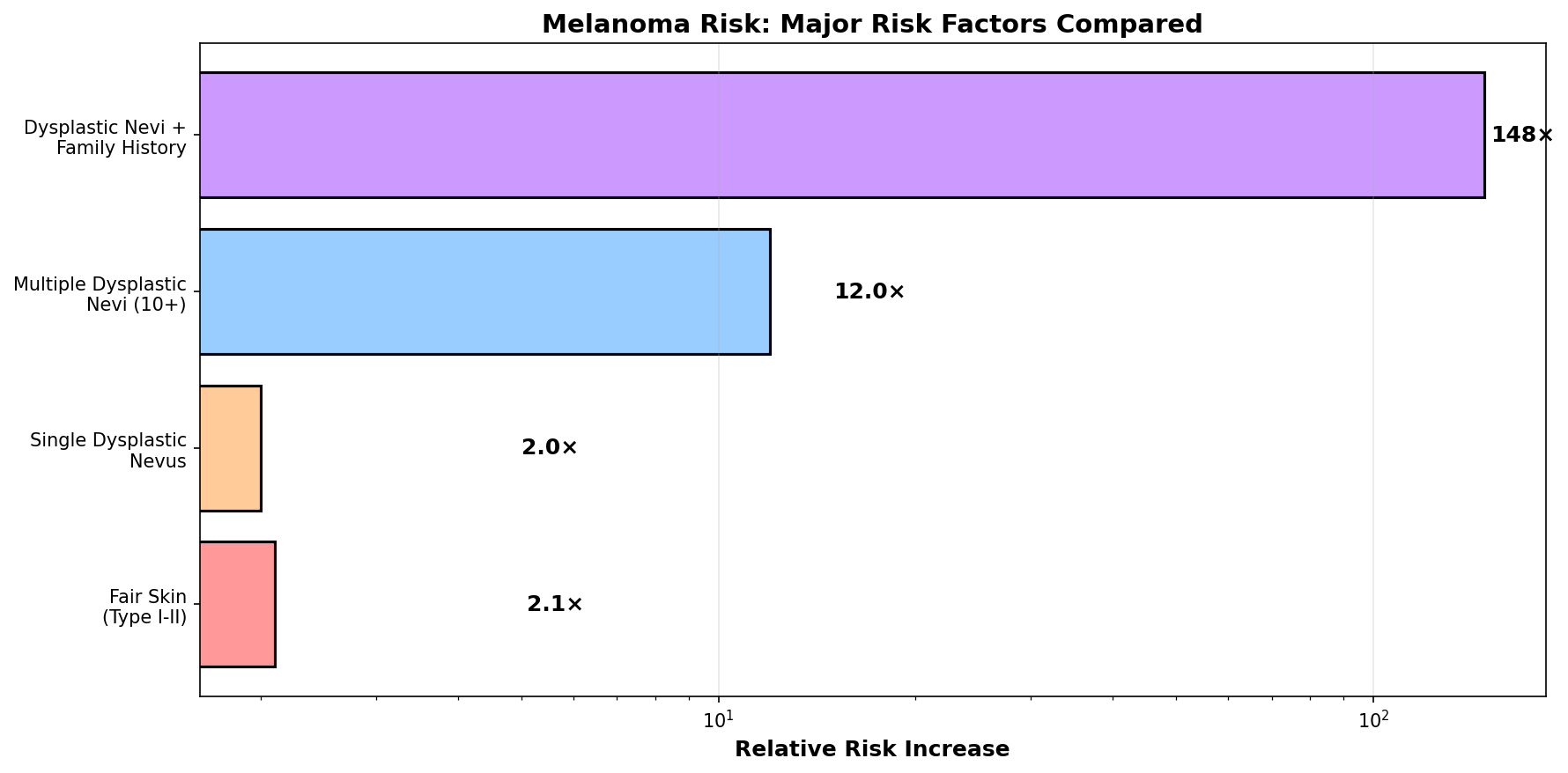
Figure 3: Individual ABCDE criteria appear at varying frequencies in melanoma. Colour and texture variation appears in 94% of melanomas, while asymmetry appears in only 46.3% of small lesions. This highlights why no single criterion is diagnostic.
One powerful concept is the "ugly duckling sign". If a mole looks notably different from your other moles, even if it does not meet all ABCDE criteria, that dissimilarity warrants evaluation. Many patients do not have just one mole, they may have more and sometimes the concerning one stands out precisely because it breaks from the usual pattern.
Dermatologists also use dermoscopy, a handheld magnifying device called a dermatoscope, that shows subsurface features invisible to the naked eye. With proper training, dermoscopy increases diagnostic accuracy to 84-91.6% sensitivity. However, this benefit requires expertise and explains why specialists catch lesions that primary care doctors miss.
Important Limitations of ABCDE
The ABCDE rule catches approximately 47–83% of melanomas, depending on the study and how they are applied. This means it will occasionally miss melanomas, especially:
- Early or flat lesions without classic features
- Small lesions under 6mm
- Amelanotic (non-pigmented) melanomas
- In-situ melanomas (confined to the superficial layer)
- Special presentations like lentigo maligna or desmoplastic melanoma
Additionally, benign moles can meet several ABCDE criteria, generating false alarms. For example, atypical or dysplastic nevi (distinct from melanoma but requiring monitoring) often appear asymmetrical with colour variation and irregular borders.
Who is at a High Risk: Understanding Your Personal Factors
Risk for skin cancer varies significantly based on genetics, sun exposure history, skin type and medical status.
Skin Phototype: The Foundation of Risk
Fair-skinned individuals (Skin Types I–II) face a 2.1 times higher melanoma risk compared to darker skin types. This is because fair skin produces less protective melanin, rendering it more vulnerable to UV damage. Fair skin is particularly associated with invasive melanomas rather than surface-confined disease. However, this does not mean darker-skinned people are immune. Melanoma occurs across all races. However, diagnosis is often delayed in people of colour because the disease may appear in less obvious locations, such as the palms, soles and nail beds, and present with different clinical features than in fair-skinned populations. This contributes to worse outcomes in some demographic groups.
Genetic and Family Factors
Melanoma can be inherited. If one or both parents developed melanoma, the risk increases substantially. Having multiple family members with melanoma pushes the risk even higher. Perhaps most important is the presence of dysplastic, atypical nevi, moles with irregular borders, multiple colours and larger size, that suggest genetic predisposition. A person with many dysplastic nevi faces a 47 times higher risk of melanoma compared to someone without, and if that person also has a family history of melanoma, the risk is 148 times higher.
Specific genes also carry a high risk. CDKN2A mutations cause hereditary melanoma, while variations in MC1R (the gene regulating melanin production) increase risk among carriers. These genetic factors explain why some families experience multiple melanoma diagnoses across generations.
Prior Sun Exposure and Skin Damage
UV radiation accounts for 63–90% of melanoma cases. Intense, intermittent sun exposure (vacations, sunburns, weekend beach trips) carries greater melanoma risk. Cumulative chronic exposure (occupational outdoor work) more strongly predicts non-melanoma skin cancers.
Childhood sunburns carry particular weight. Early-life sun exposure correlates most strongly with adult melanoma development. If you received significant sunburns before age 18, you carry elevated lifelong risk.
Visible signs of cumulative sun damage such as age spots, rough scaly patches (actinic keratoses) and leathery skin texture, indicate substantial UV dose and warrant professional screening regardless of personal cancer history.

Figure 4: Atypical nevi (dysplastic moles) present the highest clinical risk factor for melanoma (47 times increased risk), followed by family history of melanoma (7–10 times increased risk), fair skin phototype (2.1 times increased risk) and prior sun exposure patterns.
Indoor Tanning: An Underestimated Risk
Indoor tanning creates a dose-dependent risk: the more frequently people use tanning beds, especially starting in adolescence, the higher their melanoma risk. Regular indoor tanners also show concerning behavioural patterns: higher sunburn rates, avoidance of sun protection and lower rates of skin cancer screening. This suggests indoor tanning serves as a marker of overall risk-taking behaviour around sun exposure.
Restricting indoor tanning access for minors under 18 could prevent melanoma incidence and mortality while saving millions in treatment costs.
Immunosuppression
Organ transplant recipients on chronic immunosuppressive therapy face elevated skin cancer risk. Interestingly, melanoma becomes the most common non-keratinocyte skin cancer (61.8% of cases) in this population, which differs from general population patterns where non-melanoma skin cancers dominate.
Recent research shows switching from calcineurin inhibitor-based immunosuppression to mTOR inhibitors dramatically reduces skin cancer development and occasionally causes regression of existing lesions. If you are a transplant recipient, discuss immunosuppressive regimen optimisation with the transplant team.
Demographic Disparities: Some People Face Greater Challenges
Skin cancer does not affect all populations equally. Caucasians experience melanoma at 24.6 per 100,000, compared to 0.8 per 100,000 for African Americans, a 30-fold disparity.
This gap reflects both biological differences (melanin production) and healthcare access disparities. Darker-skinned individuals often receive delayed diagnoses because melanoma presentations differ and awareness campaigns typically feature light-skinned patients. Additionally, unequal access to dermatology care and preventive screening contributes to later-stage disease at diagnosis and worse survival outcomes.
Median age at diagnosis also varies: 59–63 years for caucasian and African American patients versus 52–56 years for Hispanic, American Indian and Asian/Pacific Islander populations.

Figure 5: Melanoma incidence varies dramatically by race and ethnicity in the United States. Caucasians experience 30-fold higher incidence than African Americans, reflecting both biological and healthcare access factors.
Survival: Why Stage at Detection Matters
The survival statistics reveal that detecting melanoma early transforms prognosis.
- Localised melanomas (confined to skin): 99.4% five-year survival
- Regional melanomas (spread to nearby lymph nodes): 64% five-year survival
- Distant melanomas (spread to other organs): 35.6% five-year survival
This 63.8% point difference between localised and distant disease illustrates why catching melanoma before it spreads is the most important factor determining outcomes. A person diagnosed with localised melanoma at age 50 has vastly better prospects than someone with the same disease diagnosed 5 years later at stage III or IV.
Research on screening programs confirms this. Serial surveillance programs that examined patients every 6 months detected 100% of melanomas at an early stage. All lesions were either in situ (non-invasive) or in the radial growth phase with minimal thickness. Over 17 years of follow-up, this screening protocol resulted in zero melanoma deaths, metastases and recurrences.
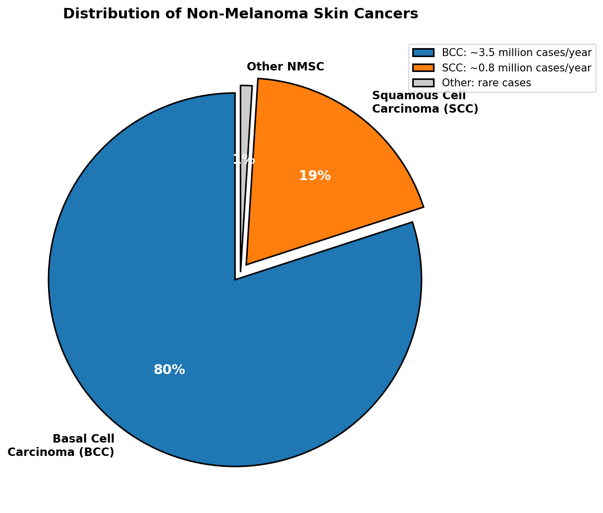
Figure 6: Five-year survival rates differ dramatically by stage at diagnosis. Early detection through screening programs like ABCDE achieves 99.4% survival in localised disease, compared to 35.6% when cancer has spread distantly.
When to Seek Professional Evaluation
If you meet any of these criteria, seek professional skin examination:
- A mole meets two or more ABCDE criteria
- A mole has changed noticeably in recent months
- A mole looks distinctly different from the other moles
- Symptoms include itching, tenderness, bleeding or scabbing
- A lesion has not healed after 4 weeks
- There are multiple dysplastic nevi
- A family history of melanoma
Screening Frequency Recommendations
High-risk patients (age 50+, personal history of melanoma or non-melanoma skin cancer, family history, multiple dysplastic nevi or immunosuppression) should have annual professional dermatology screening.
Lower-risk patients benefit more from self-examination and awareness of ABCDE changes. Expensive frequent screening is not evidence-based for average-risk individuals, though annual checks after age 50 remain reasonable. Regardless of risk level, regular self-examination catches many melanomas that would otherwise be missed.
Prevention: What Actually Reduces Your Risk
Protective Clothing
Research consistently identifies protective clothing as the most reliable photoprotection strategy. Unlike sunscreen, which relies on correct application and reapplication, clothing provides passive, foolproof protection.
Wear protective clothing: long-sleeved shirts, trousers and broad-brimmed hats (>3 inches). Modern sun-protective fabrics with UPF ratings are lightweight, breathable and fashionable. Tightly-woven, dark-coloured fabrics offer better protection than standard summer clothing.
Sunscreen
Sunscreen provides helpful supplementary protection when applied correctly using SPF 30+ broad-spectrum (protecting against both UVA and UVB) that is applied generously and reapplied every 2 hours or after swimming or exercise. Most people apply too little sunscreen, skip reapplication and may compensate by spending more time in the sun. Sunscreen works best as part of a comprehensive strategy, not as a standalone solution.
Behaviour
- Avoid indoor tanning completely, the evidence against it is consistent and substantial
- Plan outdoor activities for early morning or late afternoon
- Avoid peak UV hours (10am to 4pm) when possible
- Use umbrellas or seek shade during peak UV hours
Patient Education
Intensive education programs demonstrably improve sun-protective behaviour. Knowledge improvements and behavioural changes persist months after intervention. The most effective programs combine structured education, counseling, repeated reinforcement and personalised risk assessment.
What It Means to Be a Melanoma Survivor
For those successfully treated after early detection of melanoma, the prognosis is generally positive, however, the psychological impact extends far beyond physical healing.
Approximately 31% of melanoma survivors report anxiety or depression and fear of recurrence persists for years. Many report unmet information needs even years after treatment. Those treated for advanced melanoma with immunotherapy experience significant treatment toxicity, 92% experienced immune checkpoint inhibitor side effects with 43% experiencing severe toxicity requiring steroid treatment. Survivors also report greater physical fatigue, financial difficulties and lower overall quality of life scores compared to age-matched controls.
This underscores a critical point, prevention is vastly preferable to treatment and survival. Avoiding skin cancer through early detection and protective measures is far better than living with the long-term burden of melanoma, even when physically cured.
Putting It All Together: Your Personal Action Plan
Here is how to translate the ABCDE framework into practical action:
First, know your risk level:
- Fair skin, multiple moles, family history of melanoma or visible sun damage = higher risk
- Transplant recipient or immunosuppressed = higher risk
- Minimal moles, dark skin and minimal sun exposure = lower risk (but not zero risk)
Second, establish a routine:
- Once a month, examine your entire body checking for new lesions and changes to existing ones
- If you are at a high risk, arrange annual professional screening
- Know the ABCDE criteria, but remember they are guidelines
Third, prioritise prevention:
- Make protective clothing your cornerstone strategy
- Avoid peak sun hours when possible
- Use sunscreen correctly as supplementary protection
- Avoid indoor tanning entirely
Fourth, act on concerning findings:
- Any moles meeting 2+ ABCDE criteria warrants professional evaluation
- Any change over weeks to months warrants evaluation
- Any lesion that looks notably different from your other moles warrants evaluation
- When in doubt, get it checked
The ABCDE framework works because it is simple, memorable and based on biological differences between benign and malignant lesions. It is not perfect, but has been refined over 40 years of clinical use and has saved countless lives by encouraging early detection.
Further Reading
Understanding Melanoma and Skin Cancer Basics
- What does melanoma look like? Recognising the ABCDEs
- Moles, dysplastic nevi, and the risk of melanoma
- Melanoma: symptoms, staging, and treatment overview
- How to perform a skin self-examination
- A visual guide to melanoma diagnosis and features
Risk Factors and Prevention
- Reducing skin cancer risk: sun exposure and protective strategies
- Ultraviolet (UV) radiation and skin cancer risk
- Sunscreen safety and efficacy for melanoma prevention
- Indoor tanning and melanoma risk
- Screening and early detection in high-risk populations
Professional Clinical Resources
- Melanoma diagnosis and clinical decision-making
- Dermoscopy and advanced diagnostic techniques
- Atypical mole syndrome and hereditary melanoma
References/Helpful Resources
- National Cancer Institute. What does melanoma look like? [Internet]. [cited 2025 June 1]. Available from: https://www.cancer.gov/types/skin/melanoma-photos
- National Cancer Institute. Common moles, dysplastic nevi, and risk of melanoma [Internet]. [cited 2025 June 1]. Available from: https://www.cancer.gov/types/skin/moles-fact-sheet
- Cleveland Clinic. ABCDEs of melanoma: warning signs of skin cancer [Internet]. [cited 2025 June 1]. Available from: https://my.clevelandclinic.org/health/diagnostics/8648-skin-self-exam
- Cleveland Clinic. Melanoma: symptoms, staging and treatment [Internet]. [cited 2025 June 1]. Available from: https://my.clevelandclinic.org/health/diseases/14391-melanoma
- Mayo Clinic. Melanoma pictures: helpful images for identification [Internet]. [cited 2025 June 1]. Available from: https://www.mayoclinic.org/diseases-conditions/melanoma/in-depth/melanoma/art-20546856
- Mayo Clinic. Melanoma diagnosis and treatment overview [Internet]. [cited 2025 June 1]. Available from: https://www.mayoclinic.org/diseases-conditions/melanoma/diagnosis-treatment/drc-20374889
- National Cancer Institute. Moles to melanoma: recognising the ABCDE features [Internet]. [cited 2025 June 1]. Available from: https://moles-melanoma-tool.cancer.gov/
- Ferris LK, Harkes JA, Gilbert S, et al. Early detection of melanoma: reviewing the ABCDEs. J Am Acad Dermatol. 2015;72(5):717-723. PubMed: 25698455.
- Rigel DS, Friedman RJ, Kopf AW, et al. The clinical diagnosis of early malignant melanoma: expansion of the ABCD criteria to improve diagnostic sensitivity. Arch Dermatol. 2000;136(4):451-458. PubMed: 10673456.
- Kimura T, Takeuchi M, Kanda Y, et al. The dermoscopic (7FFM) versus the clinical (ABCDE) diagnosis of small diameter melanoma. Br J Dermatol. 2000;143(5):977-981. PubMed: 10846255.
- Scope A, Benvenuto-Andrade C, Agero AL, et al. Reproducibility of the clinical criteria (ABCDE rule) and dermatoscopic features (7FFM) for the diagnosis of malignant melanoma. Arch Dermatol. 2004;140(10):1220-1225. PubMed: 11358731.
- National Institute of Health, National Cancer Institute. Trends in melanoma incidence, prevalence, stage at diagnosis, and survival. Cancer Epidemiol Biomarkers Prev. 2023;32(4):445-451. PMC: 11529802.
- Garbe C, Leiter U, Ellwanger U, et al. Epidemiology of melanoma. NCBI Bookshelf: Cutaneous Melanoma. 2021. NBK481862.
- Sánchez MC, Fabi SG, Wu MT, et al. The impact of melanoma imaging biomarker cues on detection sensitivity and specificity in melanoma versus clinically atypical nevi. Dermatol Surg. 2024;50(9):e313-e318. PMC: 11394255.
- Pediatric melanoma consortium. Pediatric melanoma: results of a large cohort study and proposal for modified ABCD detection criteria for children. J Am Acad Dermatol. 2013;68(6):913-925. PubMed: 23395590.
- CDC. Vital signs: melanoma incidence and mortality trends and projections, United States, 1982–2030. MMWR Morb Mortal Wkly Rep. 2015;64(21):591-596. Available from: https://www.cdc.gov/mmwr/preview/mmwrhtml/mm6421a6.htm
- Epidemiology of melanoma. NCBI Bookshelf. Available from: https://www.ncbi.nlm.nih.gov/books/NBK481862/
- Patterns and trends in melanoma mortality in the United States, 1999–2020. PMC. 2023;11221171. Available from: https://www.ncbi.nlm.nih.gov/pmc/articles/PMC11221171/
- Non-melanoma skin cancer epidemiology. NCBI Bookshelf. The incidence and clinical analysis of non-melanoma skin cancer. PMC. 2021;7900109. Available from: https://www.ncbi.nlm.nih.gov/pmc/articles/PMC7900109/
- Jemal A, Saraiya M, Wingo PA, et al. Racial and ethnic variations in incidence and survival of cutaneous melanoma in the United States, 1999-2006. J Am Acad Dermatol. 2012;66(4):e119-e131. PubMed: 22018064.
- Diepgen TL, Mahler V. The epidemiology of skin cancer. Br J Dermatol. 2002;146(Suppl 61):1-6. PMC: 2010011.
- Epidemiology and primary location of melanoma in Asian patients. PMC. 2023;11127025. Available from: https://www.ncbi.nlm.nih.gov/pmc/articles/PMC11127025/
- Global epidemiological trends in the incidence and mortality for melanoma. PubMed. 2024;40125001. Available from: https://pubmed.ncbi.nlm.nih.gov/40125001/
- Global burden of cutaneous melanoma in 2020 and projections to 2040. PubMed. 2022;35353115. Available from: https://pubmed.ncbi.nlm.nih.gov/35353115/
- Spatiotemporal trends of the global burden of melanoma in 204 countries and territories from 1990 to 2019. PubMed. 2021;34872041. Available from: https://pubmed.ncbi.nlm.nih.gov/34872041/
- Basal cell carcinoma. StatPearls. NCBI Bookshelf. Available from: https://www.ncbi.nlm.nih.gov/books/NBK482439/
- Incidence and trends of basal cell carcinoma and cutaneous squamous cell carcinoma in Olmsted County, Minnesota, 2000 to 2010. PMC. 2017;5535132. Available from: https://www.ncbi.nlm.nih.gov/pmc/articles/PMC5535132/
- Trends in basal cell carcinoma incidence rates: a 16-year analysis. PMC. 2019;5872246. Available from: https://www.ncbi.nlm.nih.gov/pmc/articles/PMC5872246/
- Epidemiology of basal and cutaneous squamous cell carcinoma in the UK 2013–15. PMC. 2020;7379277. Available from: https://www.ncbi.nlm.nih.gov/pmc/articles/PMC7379277/
- Early detection of melanoma improves survival. PubMed. 2009;19938560. Available from: https://pubmed.ncbi.nlm.nih.gov/19938560/
- Serial screening for melanoma: measures and strategies that have consistently achieved early detection and cure. PubMed. 2011;21369639. Available from: https://pubmed.ncbi.nlm.nih.gov/21369639/
- Screening, early detection, education, and trends for melanoma: current status (2007-2013) and future directions. PubMed. 2014;25219716. Available from: https://pubmed.ncbi.nlm.nih.gov/25219716/
- Melanoma risk factors and genetic risk. PubMed. 2021;23753. Available from: https://pubmed.ncbi.nlm.nih.gov/21923753/
- Genetic risk factors in melanoma etiopathogenesis and the role of genetic counseling. PMC. 2022;9519167. Available from: https://www.ncbi.nlm.nih.gov/pmc/articles/PMC9519167/
- Epidemiological trends in skin cancer. PMC. 2015;5424654. Available from: https://pmc.ncbi.nlm.nih.gov/articles/PMC5424654/
- Global burden of skin cancer and its subtypes. PMC. 2024;12443545. Available from: https://pmc.ncbi.nlm.nih.gov/articles/PMC12443545/
- Global, regional and national incidence, mortality and disability-adjusted life-years of skin cancers. PMC. 2021;8290243. Available from: https://www.ncbi.nlm.nih.gov/pmc/articles/PMC8290243/
- The role of sunscreen in protection from melanoma. PubMed. 2024;40581419. Available from: https://pubmed.ncbi.nlm.nih.gov/40581419/
- Sunscreen safety and efficacy for the prevention of cutaneous neoplasm. PMC. 2023;11022667. Available from: https://www.ncbi.nlm.nih.gov/pmc/articles/PMC11022667/
- Risk factors for melanoma incidence in prospective follow-up: the importance of atypical dysplastic nevi. PubMed. 1994;8053695. Available from: https://pubmed.ncbi.nlm.nih.gov/8053695/
- Clinically recognized dysplastic nevi: a central risk factor for cutaneous melanoma. PubMed. 1997;9145715. Available from: https://pubmed.ncbi.nlm.nih.gov/9145715/
- Atypical mole syndrome and dysplastic nevi: identification of populations at risk for developing melanoma. PubMed. 2011;21552679. Available from: https://pubmed.ncbi.nlm.nih.gov/21552679/
- Cutaneous melanoma more likely to be invasive in fairer skin phototypes: a retrospective observational study. PubMed. 2021;34526202. Available from: https://pubmed.ncbi.nlm.nih.gov/34526202/
- Skin colour and skin cancer: MC1R, the genetic link. PubMed. 2002;12394181. Available from: https://pubmed.ncbi.nlm.nih.gov/12394181/
- Eye and hair colour, skin type and constitutive skin pigmentation as risk factors for basal cell carcinoma and cutaneous malignant melanoma. PubMed. 2000;10086866. Available from: https://pubmed.ncbi.nlm.nih.gov/10086866/
- Skin cancer risk characterized for solid organ transplant recipients. National Cancer Institute. Available from: https://dceg.cancer.gov/news-events/news/2022/organ-transplant-skin-cancer
- Immunosuppressive regimens and skin cancer risk in solid-organ transplant recipients. PubMed. 2023;41308073. Available from: https://pubmed.ncbi.nlm.nih.gov/41308073/
- Azathioprine increases the risk of non-melanoma skin cancer among organ transplant recipients. PMC. 2024;12890441. Available from: https://www.ncbi.nlm.nih.gov/pmc/articles/PMC12890441/
- Photoprotection prevents skin cancer: let's make it fashionable to wear sun-protective clothing. PubMed. 2017;28319625. Available from: https://pubmed.ncbi.nlm.nih.gov/28319625/
- Preventing skin cancer: findings of the Task Force on Community Preventive Services on reducing exposure to ultraviolet light. PubMed. 2003;14561953. Available from: https://pubmed.ncbi.nlm.nih.gov/14561953/
- Slip versus slop: a head-to-head comparison of UV-protective clothing to sunscreen. PMC. 2022;8833350. Available from: https://www.ncbi.nlm.nih.gov/pmc/articles/PMC8833350/
- Surveillance of patients for early detection of melanoma: patterns in dermatologist vs patient discovery. PubMed. 2011;21690529. Available from: https://pubmed.ncbi.nlm.nih.gov/21690529/
- Skin photodamage and melanomagenesis: a comprehensive review. PMC. 2024;12153675. Available from: https://www.ncbi.nlm.nih.gov/pmc/articles/PMC12153675/
- Cumulative sun exposure and melanoma in a population-based case-control study. PubMed. 2022;35205756. Available from: https://pubmed.ncbi.nlm.nih.gov/35205756/
- Association of indoor tanning frequency with risky sun protection practices and skin cancer screening. PubMed. 2016;27732686. Available from: https://pubmed.ncbi.nlm.nih.gov/27732686/
- Melanoma prevention: avoiding indoor tanning and minimizing overexposure to the sun. PubMed. 2016;27824997. Available from: https://pubmed.ncbi.nlm.nih.gov/27824997/
- Indoor tanning and the risk of overall and early-onset melanoma and non-melanoma skin cancer: systematic review and meta-analysis. PMC. 2021;8656707. Available from: https://www.ncbi.nlm.nih.gov/pmc/articles/PMC8656707/
- Patient-reported outcomes in melanoma survivors at 1, 3 and 5 years post-diagnosis. PubMed. 2020;32140919. Available from: https://pubmed.ncbi.nlm.nih.gov/32140919/
- Exploring the melanoma survivorship experience: a qualitative study. PubMed. 2020;33544884. Available from: https://pubmed.ncbi.nlm.nih.gov/33544884/
- Comparison of quality of life among long-term melanoma survivors and non-melanoma controls. PubMed. 2018;28243994. Available from: https://pubmed.ncbi.nlm.nih.gov/28243994/
- Depression, anxiety and quality of life in long-term survivors of malignant melanoma. PMC. 2015;4304781. Available from: https://www.ncbi.nlm.nih.gov/pmc/articles/PMC4304781/
- Psychosocial care needs of melanoma survivors: are they being met? PubMed. 2015;26296089. Available from: https://pubmed.ncbi.nlm.nih.gov/26296089/
- Educational outcomes regarding skin cancer in organ transplant recipients. PubMed. 2006;16785373. Available from: https://pubmed.ncbi.nlm.nih.gov/16785373/
- Successful behavioral interventions to prevent cancer: the example of skin cancer. PubMed. 1996;9611622. Available from: https://pubmed.ncbi.nlm.nih.gov/9611622/
- A multimodal skin cancer prevention program for heart transplant patients. PubMed. 2019;29940790. Available from: https://pubmed.ncbi.nlm.nih.gov/29940790/
