

For most women, a mammogram every few years is a sensible and sufficient screening strategy. But for women carrying certain genetic mutations, such as changes in the BRCA1 or BRCA2 genes, or for those with a strong family history of breast cancer, a mammogram alone leaves too much to chance. It can miss more than half of cancers in this group. That is not a minor statistical shortcoming; it is the difference between finding a small, curable tumour and discovering one that has already spread.
Breast MRI (magnetic resonance imaging) was introduced into high-risk surveillance programmes precisely to close that gap. Over two decades of evidence now show it dramatically improves early detection and, critically, improves survival. This article explains who needs MRI surveillance, how the scan works, what the benefits and trade-offs look like, and what current guidelines say on both sides of the Atlantic.
Who Counts as High-Risk?
Before exploring MRI itself, it is worth understanding what "high risk" actually means. For the average woman, the lifetime risk of developing breast cancer is around 12%: roughly 1 in 8 over a lifetime. That is already a significant probability, but it is the baseline against which elevated risk is measured.
The groups most clearly identified as high-risk are:
- BRCA1 mutation carriers: estimated lifetime breast cancer risk of 60–79.5% (roughly 6 in 8 women)
- BRCA2 mutation carriers: estimated lifetime risk of 63–88%
- PALB2 mutation carriers: lifetime risk of 40–60%
- TP53 mutation carriers (Li-Fraumeni syndrome): lifetime risk above 40%
- ATM mutation carriers: lifetime risk of 20–40%
- Women with prior chest radiotherapy between the ages of 10 and 30 (for example, Hodgkin lymphoma survivors): cumulative breast cancer risk of 35% by age 50, which exceeds even some BRCA1 figures at that age
- Women with no confirmed genetic mutation but a calculated lifetime risk of 20% or higher based on family history
The chart below shows how dramatically lifetime risk rises across different genetic mutations, compared with the 12% background rate.
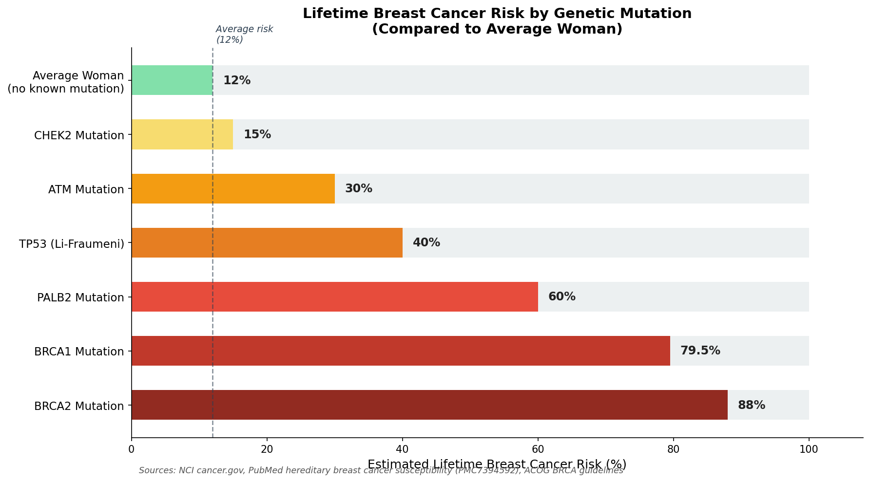 Figure 1: Estimated lifetime breast cancer risk varies enormously by genetic mutation. Data from NCI cancer.gov, PMC hereditary breast cancer susceptibility studies, and ACOG BRCA guidelines.
BRCA1 and BRCA2 mutations are rarer than many people realise: approximately 1 in 300–400 people in the general population carry one. However, rates are notably higher in people of Ashkenazi Jewish descent, where roughly 1 in 40 carry one of three founder mutations. Genetic testing, offered following a referral to a clinical genetics service, can identify these mutations before cancer develops.
It is also worth noting that some women discover elevated risk not through genetic testing but through a previous biopsy. Women diagnosed with lobular carcinoma in situ (LCIS) carry approximately nine times the average breast cancer risk, while atypical ductal hyperplasia (ADH) carries four to five times the average risk. A biopsy finding like this does not mean cancer is present now; it is a signal to step up monitoring.
Why Mammography Falls Short for This Group
Mammography is an excellent tool for average-risk women, particularly over the age of 50. It works by detecting differences in tissue density and can spot calcifications (calcium deposits) that are often an early sign of cancer. But it has a fundamental limitation in high-risk, younger women: breast density.
Younger women tend to have denser breast tissue. On a mammogram, dense tissue appears white, and so do tumours. The result is that cancers can hide in plain sight. In high-risk women, pooled data from multiple studies show mammography detects only around 39% of cancers on average; in some studies, as few as 13%.
There is a further problem specific to BRCA1-associated cancers. They tend to grow quickly, are often hormone-receptor-negative (meaning they lack certain molecular markers that slow growth), and can reach a significant size in the interval between annual mammograms. This is not a failure of scheduling; it is a biological feature of these tumours that demands a different approach.
How Breast MRI Works
Breast MRI uses a powerful magnetic field and radio waves to create detailed three-dimensional images of breast tissue. Unlike mammography, it does not use radiation.
Women lie face-down on a padded table with openings for the breasts, which hang freely to allow imaging from all angles. Before the scan begins, a small injection of a contrast dye called gadolinium is given through a vein in the arm. This is the key step that makes MRI so effective.
Tumours grow abnormally fast and form new, poorly structured blood vessels to supply themselves. The gadolinium dye leaks into these vessels and causes the tumour to "light up" on the scan far more brightly and quickly than surrounding healthy tissue. Radiologists then study not just whether an area lights up, but how quickly and whether the enhancement fades, plateaus, or persists over time. These patterns, called kinetic curves, help distinguish cancerous tissue from benign changes.
MRI can detect cancers based on blood flow and tissue architecture rather than density alone, which is precisely why it succeeds where mammography struggles in younger, denser breasts.
What the Evidence Shows: Detection Rates
The case for MRI in high-risk surveillance rests on a substantial and consistent body of evidence. The landmark MARIBS trial, published in The Lancet in 2005, found that MRI detected 77% of tumours in high-risk women, compared with 40% for mammography. In BRCA1 carriers specifically, MRI detected 92% of cancers against just 23% for mammography. Combined, the two methods reached 94% sensitivity.
More recent meta-analyses confirm these figures. A pooled analysis covering 45,060 high-risk women found MRI's average detection rate across 11 studies reached 94% when combined with mammography, against 39% for mammography alone.
The chart below illustrates the detection sensitivity of each screening method in high-risk populations.
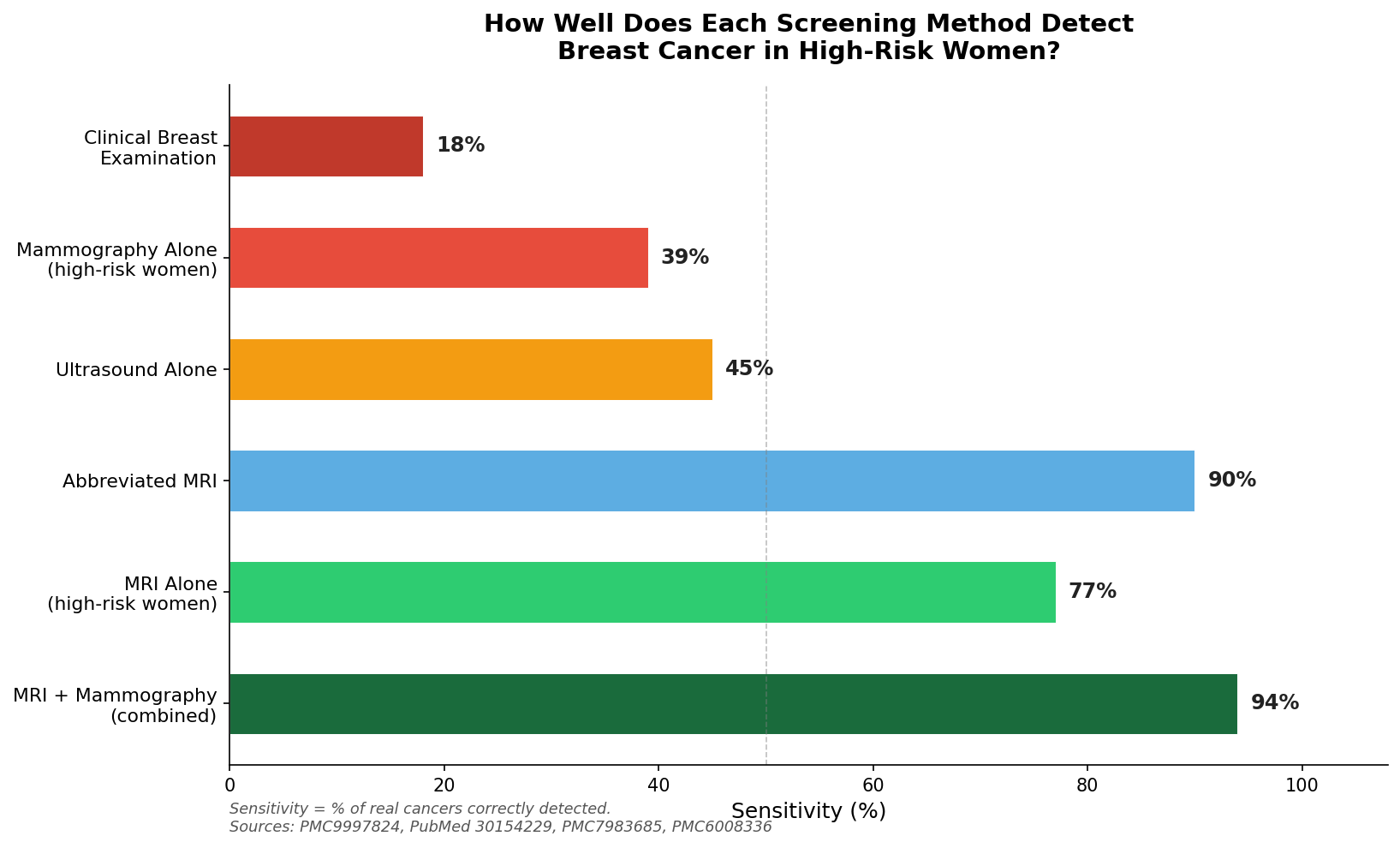 Figure 2: How well each screening method detects breast cancer in high-risk women. MRI combined with mammography achieves 94% sensitivity, compared with 39% for mammography alone. Sources: PMC9997824, PubMed 30154229, PMC7983685, PMC6008336.
In practical terms: if 100 cancers were present in a high-risk group, mammography alone would find roughly 39 of them. Adding MRI would find approximately 94. The 55 additional cancers found are not statistics; they are women whose disease can be treated while it is still small, localised, and curable.
The difference in how many cancers are actually found per 1,000 women screened is equally striking.
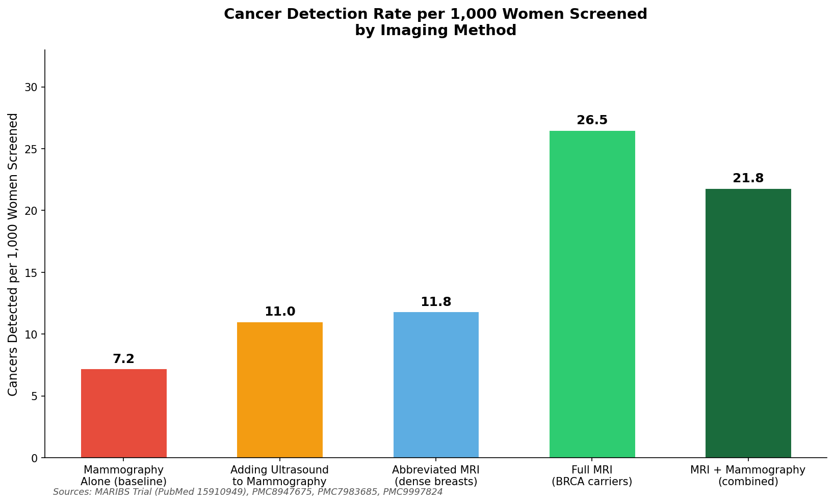 Figure 3: Cancer detection rate per 1,000 women by screening approach. MRI in BRCA carriers detects 26.5 cancers per 1,000, compared with 7.2 for mammography alone. Sources: MARIBS Trial (PubMed 15910949), PMC8947675, PMC7983685, PMC9997824.
What Stage Cancers Are Found at Matters Enormously
Detection rates are only part of the story. The stage at which a cancer is found is arguably more important, because it largely determines treatment options and prognosis.
MRI-detected cancers tend to be small and caught before they have spread to the lymph nodes (the glands under the arm that represent the next step of spread). Across multiple high-risk screening programmes, MRI-detected cancers have a median tumour size of just 8–9 mm, compared with 17 mm for tumours found by mammography or ultrasound. In one large study, the proportion of cancers with lymph node involvement at detection was 11% in the MRI group, compared with 63% in the mammography group.
In terms of formal cancer staging, 92.9% of MRI-detected cancers in high-risk programmes were classified as Stage 0 (pre-invasive) or Stage I (small and localised) in recent cohort data. This compares with around 45% at these early stages for women not receiving intensive MRI-based screening.
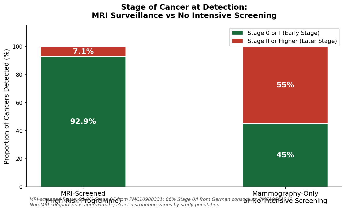 Figure 4: MRI surveillance dramatically improves the stage at which cancer is detected. Nearly 93% of MRI-detected cancers are caught at early stage (Stage 0 or I), compared with around 45% without intensive screening. Sources: PMC10988331, PMC10661242.
The Impact on Survival
The survival data from high-risk MRI surveillance programmes are among the most compelling in all of cancer screening medicine.
A major study following 1,756 BRCA1 and BRCA2 carriers over 20 years found that the risk of dying from breast cancer was 3.2% in the MRI surveillance group, compared with 14.9% in women without MRI surveillance. That is a more than fourfold difference in the likelihood of dying from this disease over two decades.
A broader study across 59 centres in 11 countries (involving 2,488 women) found that BRCA1 carriers receiving annual MRI had a hazard ratio for breast cancer mortality of just 0.20 compared with those not receiving MRI surveillance: meaning their risk of dying from breast cancer was roughly five times lower.
The chart below shows three key survival metrics comparing women with and without annual MRI surveillance.
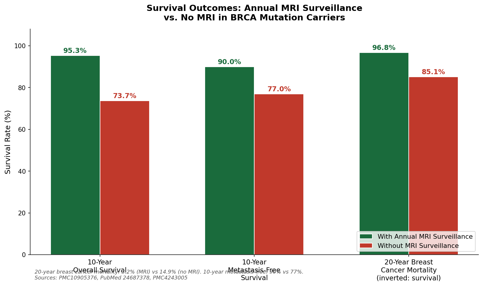 Figure 5: Survival outcomes in BRCA mutation carriers with and without annual MRI surveillance. Data from PMC10905376, PubMed 24687378, PMC4243005.
A 10-year metastasis-free survival of 90% in the MRI group versus 77% in the non-MRI group means that, among 100 women with BRCA mutations, 13 more reach the ten-year mark free of any cancer spread when they receive annual MRI. The difference in the 20-year overall survival figures is equally striking: 95.3% versus 73.7%.
These figures represent real people, not abstractions. For women who know they carry a BRCA mutation and are weighing whether to commit to annual surveillance, these numbers are among the most important in the literature.
The Trade-Off: False Positives and Unnecessary Procedures
Honest reporting of the evidence requires acknowledging MRI's most significant limitation: it produces a considerably higher rate of false-positive results than mammography. A false positive is a finding that triggers further investigation (another scan, a biopsy) but ultimately turns out not to be cancer.
In high-risk surveillance cohorts, the false-positive rate for MRI is approximately 23%, compared with roughly 2% for mammography alone. The callback rate for follow-up imaging is 14.8% with MRI versus 1.6% with mammography. Of all MRI-guided biopsies performed, only around 44.8% find cancer; the other 55.2% return a benign result.
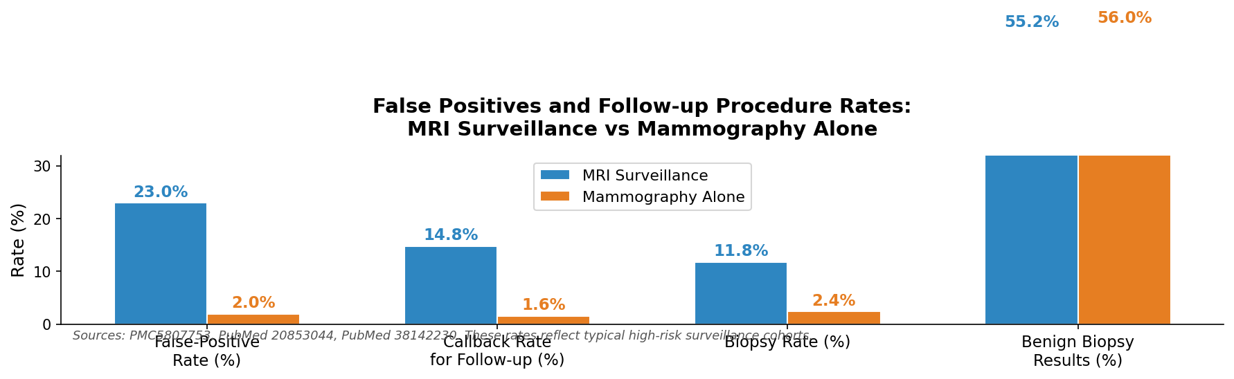 Figure 6: MRI surveillance leads to significantly higher rates of false positives, callbacks, and benign biopsy results compared with mammography alone. Sources: PMC5807753, PubMed 20853044, PubMed 38142230.
This is not a trivial issue. Waiting for biopsy results causes real anxiety, and some women find the follow-up process distressing. Studies show that around 37% of women experience anxiety during the MRI scan itself. However, the same research consistently finds that overall quality of life for women in MRI surveillance programmes is not worse than for those receiving standard mammography, and most women in high-risk programmes report feeling more supported and better cared for by their surveillance team than those on standard pathways.
The critical context is this: for a woman with a BRCA mutation or a 20%+ lifetime risk, the balance of benefit versus harm is firmly in favour of MRI. The survival data described above reflect a group that accepted some additional callbacks and biopsies in exchange for a dramatically higher probability of catching cancer early. At lower levels of risk, this trade-off becomes less favourable, which is precisely why MRI is not recommended for average-risk women.
Researchers have also developed prediction models that may be able to reduce unnecessary biopsies by up to 46% without missing any additional cancers. These tools are not yet in universal clinical use, but they represent a promising direction for reducing the burden of false positives.
Practical Considerations: Contrast, Claustrophobia, and Cost
The gadolinium contrast dye used in breast MRI is very safe for most people. Severe allergic reactions occur in roughly 0.04% to 0.07% of administrations (fewer than 1 in 1,000 injections). People with severe kidney disease should not receive gadolinium because of a rare but serious risk of a condition called nephrogenic systemic fibrosis. The FDA has also identified that small amounts of gadolinium can be retained in brain tissue after multiple contrast MRI scans; no harmful effects have been identified in people with normal kidney function, but healthcare providers generally use macrocyclic gadolinium agents (the type that leaves fewer deposits) for repeated surveillance scanning, and the situation is actively monitored.
Claustrophobia is the most frequently cited barrier to MRI, affecting between 2% and 14% of people undergoing the scan. In one study, 97% of women who preferred an alternative to MRI cited claustrophobia as their reason. Management options include open-bore MRI scanners, short-acting sedation, breathing techniques, and mindfulness-based approaches. Most women who find the scan difficult can, with support, complete it successfully.
Cost is a real consideration. MRI is significantly more expensive than mammography. However, studies of cost-effectiveness for high-risk groups consistently find it represents good value by standard healthcare metrics. For BRCA1 and BRCA2 carriers, the cost per quality-adjusted life year (QALY) gained by adding MRI to mammography has been estimated at $25,277 to $50,900 in US studies. The standard threshold used in US healthcare is $50,000 to $100,000 per QALY, placing MRI for this group well within acceptable limits. In UK analyses, the cost per cancer detected through MRI in BRCA1 carriers was approximately £11,731: a figure that compares favourably with many other funded screening interventions. As with the clinical benefits, cost-effectiveness becomes less favourable when MRI is applied to lower-risk groups, reinforcing why the guidance is risk-stratified.
A shorter version of the scan, known as abbreviated breast MRI, takes just 3 to 10 minutes rather than the standard 30 to 45 minutes. Early evidence suggests it achieves nearly equivalent cancer detection rates. If this is confirmed in larger studies, abbreviated MRI could expand access considerably and reduce both cost and the burden on patients with claustrophobia or scan anxiety.
What the Guidelines Say: Who Should Have MRI, and When
Guidelines in the UK, USA, and Europe agree on the core principle: annual MRI surveillance should be offered to women with a sufficiently elevated risk of breast cancer, starting well before the ages at which most women begin any screening. The differences between guidelines mainly reflect variations in where they draw the risk threshold, which age windows they specify, and the role of mammography alongside MRI.
The chart below shows the recommended age windows for annual MRI by risk group and guideline.
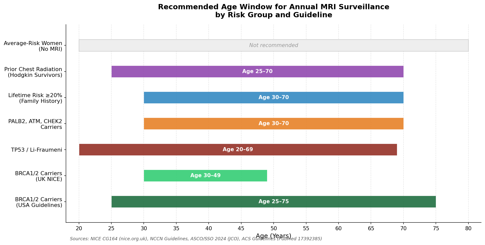 Figure 7: Annual MRI surveillance start and end ages by risk group. UK NICE guidelines cover BRCA carriers aged 30–49; US guidelines start earlier (age 25) and continue longer. Sources: NICE CG164, NCCN Guidelines, ASCO/SSO 2024, ACS Guidelines (PubMed 17392385).
In the UK (NICE Guideline CG164): annual MRI is offered to women aged 30–49 with BRCA1, BRCA2, or TP53 mutations, and to women aged 30–49 with a lifetime risk of 20% or higher. Women with TP53 (Li-Fraumeni syndrome) are offered MRI from age 20. NICE does not currently recommend MRI for BRCA carriers aged 50 and over (citing a less favourable benefit-to-harm ratio in older women, whose breast tissue is typically less dense and for whom mammography works better). The NHS provides equitable access to MRI and annual mammography for the highest-risk groups, based on evidence from the MARIBS trial published in The Lancet.
In the USA (American Cancer Society, NCCN, and ASCO/SSO 2024 guidelines): annual MRI is recommended from age 25 for BRCA1 and BRCA2 carriers, with annual mammography added from age 30. The USA starts both surveillance and combined imaging earlier than UK guidelines, and there is no fixed upper age limit. A 2024 NCCN update introduced a new, lower-risk category: women with a five-year AI-estimated breast cancer risk of 1.7% or higher may be considered for supplemental imaging including MRI or ultrasound, representing a significant broadening of the surveillance net.
In Europe (European Society of Breast Imaging): the guidance broadly aligns with US recommendations for genetics-based high risk, recommending annual MRI from age 25–30 for women with BRCA or equivalent mutations, with annual mammography added from age 30 or 40 depending on specific risk.
The table below summarises the conditions that qualify women for MRI surveillance under major guidelines:
| Risk Category | Typical MRI Start Age |
|---|---|
| BRCA1 or BRCA2 mutation (UK NICE) | Age 30 |
| BRCA1 or BRCA2 mutation (US guidelines) | Age 25 |
| TP53 / Li-Fraumeni syndrome | Age 20 |
| Prior chest radiation aged 10–30 | Age 25 or 8 years post-treatment (whichever is later) |
| PALB2, ATM, or CHEK2 mutations | Age 30–40 (depending on risk level) |
| Lifetime risk 20% or higher (family history) | Age 30–35 |
| LCIS or atypical hyperplasia on prior biopsy | Age 30+ (case-by-case assessment) |
Women who are unsure whether they meet these criteria should speak with their GP, who can refer them for a formal risk assessment. In the UK, this is usually done through a clinical genetics service or a specialised family history clinic. In the US, genetic counsellors are typically the first point of contact for hereditary risk assessment.
What Patients Actually Experience
One concern that often arises is whether committing to annual intensive surveillance creates more anxiety than it relieves. The research on this is reassuring.
Multiple studies show that women in annual MRI surveillance programmes do not report worse health-related quality of life than women receiving standard mammography. Most women who already carry significant genetic risk find the structured surveillance programme a source of reassurance rather than alarm. Knowing that any cancer will be caught small and early is, for most, a more manageable situation than the uncertainty of less intensive monitoring.
Mammography is also consistently reported as more physically uncomfortable than MRI, despite the procedural complexities of the latter. The most difficult part of MRI for most women is the wait for results after a callback, not the scan itself.
Women who have previously had MRI scans report less anxiety at subsequent scans, which is relevant for a programme involving annual examinations over several decades.
Looking Ahead: Personalised Surveillance
Breast cancer surveillance for high-risk women is moving towards greater personalisation. Rather than following a fixed schedule based purely on which gene variant is present, clinicians are increasingly able to factor in tumour growth characteristics, other genetic markers, breast density measurements, and individual patient preferences.
Abbreviated MRI protocols offer the prospect of shorter scans with similar accuracy. Risk models using artificial intelligence are beginning to refine who falls above the 20% lifetime risk threshold with greater precision than traditional family history calculations alone.
The current evidence base is already robust enough to be confident about one thing: for women with significantly elevated genetic or familial breast cancer risk, annual MRI surveillance is one of the most effective preventive health interventions available. It finds cancers earlier, catches them at smaller sizes, and translates that advantage into substantially better long-term survival.
If you think you might be at elevated risk, the most important first step is to speak with your GP. Risk assessment and, where appropriate, genetic testing can clarify your personal situation, and the right surveillance pathway can be put in place well before cancer develops.
Further Reading
Clinical Efficacy and Detection Studies
- Meta-analysis of MRI sensitivity and specificity in high-risk women (PMC9997824)
- MARIBS Trial: MRI versus mammography in high-risk women (PubMed 15910949)
- High-risk breast MRI screening outcomes, tertiary care centre (PubMed 38142230)
- Intensified MRI surveillance in BRCA1 carriers (PubMed 30154229)
- German Consortium 10-year programme data (PMC10661242)
Survival Outcomes and Long-Term Benefits
- MRI surveillance and breast cancer mortality in BRCA1 and BRCA2 carriers (PMC10905376)
- MRI breast screening in high-risk women: cancer detection and survival analysis (PubMed 24687378)
- Survival outcomes of breast MRI screening in elevated-risk women (PubMed 32055796)
- Breast cancer mortality in BRCA carriers: MRI plus mammography programme (PMC7700272)
False Positives and Patient Experience
- False-positive diagnoses: MRI versus mammography and digital tomosynthesis (PMC5807753)
- Impact of breast cancer screening in high-risk women on health-related quality of life (PMC2364757)
- Stacked versus alternating MRI and mammography schedules and false-positive rates (PMC4340856)
Epidemiology and Risk Stratification
- BRCA1 and BRCA2: cancer risks and management (NCI PDQ)
- Hereditary breast cancer susceptibility genes (PMC7394592)
- Hodgkin lymphoma survivors and cumulative breast cancer incidence (PMC11639875)
Guidelines and Policy
- NICE CG164: Familial breast cancer recommendations
- American Cancer Society guidelines for breast screening with MRI (PubMed 17392385)
- Cancer surveillance in BRCA1/2 carriers: review of secondary prevention guidelines (PMC7322604)
- ASCO/SSO 2024 guideline on germline testing in breast cancer
Cost-Effectiveness
- Cost-effectiveness of MRI for breast cancer screening in BRCA1/2 carriers (PMC3711845)
- Cost-effectiveness of MRI versus mammography in high familial risk women (PMC2360541)
Helpful Resources
- National Institute for Health and Care Excellence. Familial breast cancer: classification, care and managing risks in people with a family history of breast cancer (CG164) [Internet]. [cited 2026 Jun 01]. Available from: https://www.nice.org.uk/guidance/cg164/chapter/recommendations
- National Health Service. Breast cancer screening [Internet]. [cited 2026 Jun 01]. Available from: https://www.nhs.uk/conditions/breast-cancer-screening/
- National Cancer Institute. BRCA1 and BRCA2: cancer risks and management (PDQ) [Internet]. [cited 2026 Jun 01]. Available from: https://www.cancer.gov/about-cancer/causes-prevention/genetics/brca-fact-sheet
- Centers for Disease Control and Prevention. Breast cancer statistics [Internet]. [cited 2026 Jun 01]. Available from: https://www.cdc.gov/breast-cancer/statistics/index.html
- Food and Drug Administration. FDA Drug Safety Communication: FDA evaluating the risk of brain deposits with repeated use of gadolinium-based contrast agents for MRI [Internet]. [cited 2026 Jun 01]. Available from: https://www.fda.gov/Drugs/DrugSafety/ucm455386.htm
- Phi XA, Houssami N, Hooning MJ, Castillo Tovar JE, Obdeijn IM, Cluze C, et al. Accuracy of screening women at familial risk of breast cancer without a BRCA1/2 mutation: individual patient data meta-analysis. Eur J Cancer. 2017;89:14–24. Available from: https://www.ncbi.nlm.nih.gov/pmc/articles/PMC9997824/
- Riedl CC, Luft N, Bernhart C, Weber M, Bernathova M, Tea MK, et al. Triple-modality screening trial for familial breast cancer underlines the importance of magnetic resonance imaging and questions the role of mammography and ultrasound regardless of patient mutation status, age, and breast density. J Clin Oncol. 2015;33(10):1128–35. Available from: https://pubmed.ncbi.nlm.nih.gov/25960720/
- Saadatmand S, Geuzinge HA, Rutgers EJ, Mann RM, de Roy van Zuidewijn DB, Zonderland HM, et al. MRI versus mammography for breast cancer screening in women with familial risk (FaMRIsc). Lancet Oncol. 2019;20(8):1136–47. Available from: https://pubmed.ncbi.nlm.nih.gov/32055796/
- Sardanelli F, Boetes C, Borisch B, Decker T, Federico M, Gilbert FJ, et al. Magnetic resonance imaging of the breast: recommendations from the EUSOMA working group. Eur J Cancer. 2010;46(8):1296–316. Available from: https://pubmed.ncbi.nlm.nih.gov/20304629/
- Heijnsdijk EA, Warner E, Gilbert FJ, Tilanus-Linthorst MM, Evans G, Causer PA, et al. Differences in natural history between breast cancers in BRCA1 and BRCA2 mutation carriers and effects of MRI screening. Cancer Epidemiol Biomarkers Prev. 2012;21(9):1616–22. Available from: https://pubmed.ncbi.nlm.nih.gov/23837641/
- Passaperuma K, Warner E, Causer PA, Hill KA, Messner S, Wong JW, et al. Long-term results of screening with magnetic resonance imaging in women with BRCA mutations. Br J Cancer. 2012;107(1):24–30. Available from: https://pubmed.ncbi.nlm.nih.gov/22669161/
- Berg WA, Zhang Z, Lehrer D, Jong RA, Pisano ED, Barr RG, et al. Detection of breast cancer with addition of annual screening ultrasound or a single screening MRI to mammography in women with elevated breast cancer risk. JAMA. 2012;307(13):1394–404. Available from: https://pubmed.ncbi.nlm.nih.gov/22474203/
- Lehman CD, Isaacs C, Schnall MD, Pisano ED, Ascher SM, Weatherall PT, et al. Cancer yield of mammography, MR, and US in high-risk women: prospective multi-institution breast cancer screening study. Radiology. 2007;244(2):381–8. Available from: https://pubmed.ncbi.nlm.nih.gov/17641362/
- Kriege M, Brekelmans CT, Boetes C, Besnard PE, Zonderland HM, Obdeijn IM, et al. Efficacy of MRI and mammography for breast-cancer screening in women with a familial or genetic predisposition. N Engl J Med. 2004;351(5):427–37. Available from: https://pubmed.ncbi.nlm.nih.gov/15282350/
- Leach MO, Boggis CR, Dixon AK, Easton DF, Eeles RA, Evans DG, et al. Screening with magnetic resonance imaging and mammography of a UK population at high familial risk of breast cancer: a prospective multicentre cohort study (MARIBS). Lancet. 2005;365(9473):1769–78. Available from: https://pubmed.ncbi.nlm.nih.gov/15910949/
- Phi XA, Saadatmand S, De Bock GH, Warner E, Sardanelli F, Leach MO, et al. Contribution of mammography to MRI screening in BRCA mutation carriers by BRCA status and age: individual patient data meta-analysis. Br J Cancer. 2016;114(6):631–7. Available from: https://pmc.ncbi.nlm.nih.gov/articles/PMC6999769/
- Emaus MJ, Bakker MF, Peeters PH, Loo CE, Mann RM, Bisschops RH, et al. MR imaging as an additional screening modality for the detection of breast cancer in women aged 50–75 years with extremely dense breasts: the DENSE trial study design. Radiology. 2015;277(2):527–37. Available from: https://pubmed.ncbi.nlm.nih.gov/26098455/
- Siu AL; US Preventive Services Task Force. Screening for breast cancer: US Preventive Services Task Force recommendation statement. Ann Intern Med. 2016;164(4):279–96. Available from: https://www.uspreventiveservicestaskforce.org/uspstf/recommendation/breast-cancer-screening
- National Cancer Institute. SEER Cancer Statistics: cancer of the breast (female) [Internet]. [cited 2026 Jun 01]. Available from: https://seer.cancer.gov/statfacts/html/breast.html
- Saslow D, Boetes C, Burke W, Harms S, Leach MO, Lehman CD, et al. American Cancer Society guidelines for breast screening with MRI as an adjunct to mammography. CA Cancer J Clin. 2007;57(2):75–89. Available from: https://pubmed.ncbi.nlm.nih.gov/17392385/
