

Overview
A ventricular septal defect (VSD) is a common congenital heart defect in infants, meaning it is present at birth in almost all children who have it. A VSD is an abnormal opening or hole in the septum (the dividing wall) that separates the two lower chambers of the heart (the left and right ventricles).
This defect allows oxygen-rich blood from the left ventricle to flow back into the right ventricle where it mixes with oxygen-poor blood travelling to the lungs, leading to various complications. Understanding VSD in babies is crucial for early detection and effective management to improve outcomes.
What is a ventricular septal defect (VSD)?
A ventricular septal defect (VSD) is essentially a hole in the heart which forms during pregnancy as your baby’s heart forms and develops. Almost all VSDs are present at birth (i.e., your baby is born with this condition).
The heart is made up of four chambers: two atria and two ventricles. The septum is a barrier which acts to separate your heart into its left and right sides. During development, the septal wall might not form fully, leaving one or more holes which can vary in size.
VSD is caused when a hole is present in the wall separating the ventricles (ventricular septal defect), whereas another congenital heart condition called atrial septal defect is caused by a hole in the wall separating the atria.
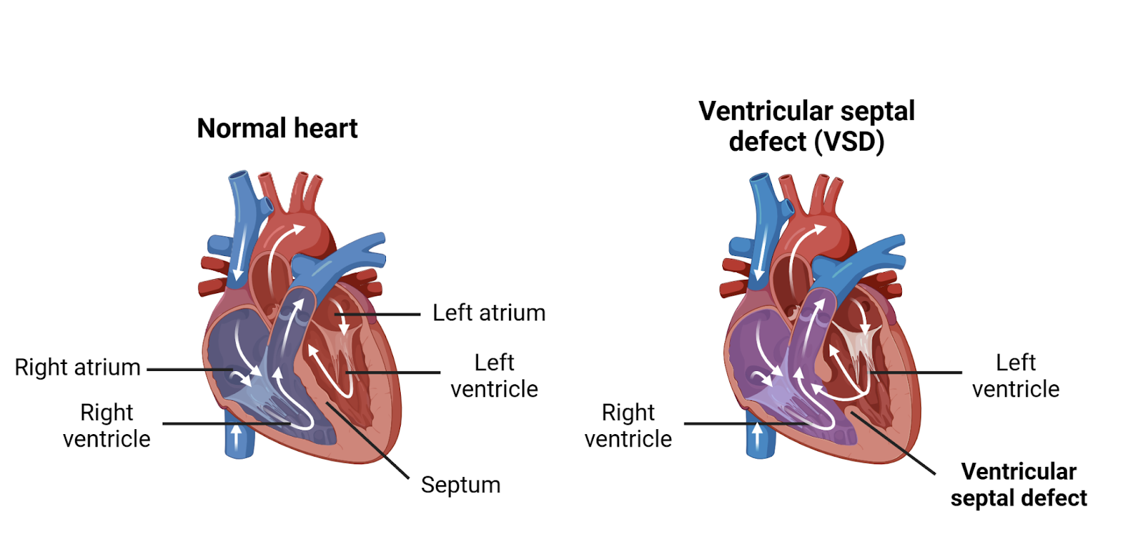
Image source: Sutherland C. Biorender.
Normally, the right ventricle pumps oxygen-poor (deoxygenated) blood into your lungs so that it can pick up oxygen. This oxygen-rich (oxygenated) blood then returns to the heart through the left side of the heart and is pumped out through the left ventricle to the rest of your body.
However, in VSD, the hole between the left and right ventricles can cause oxygen-rich blood from the left ventricle to enter and mix with oxygen-poor blood in the right ventricle. This means that extra blood is being pumped to the lungs, increasing the blood pressure in these arteries and making the heart and lungs work harder, potentially causing serious complications.1, 2
What are the different types of ventricular septal defect (VSD)?
Size of the VSD
Ventricular septal defects (VSDs) can also be classified into three different types depending on the size of the septal hole:
- Small (less than 3 mm in diameter): most VSDs fall into this classification and typically don’t cause any symptoms. Most of these cases do not require surgery as, in 85-90% of cases, the VSD will close on its own during infancy
- Moderate (3-5 mm in diameter): These VSDs often do not cause symptoms, and, if symptoms are not serious, surgery is usually delayed past infancy
- Large (6-10 mm in diameter): These large VSDs can cause serious symptoms and often require surgery before the age of 2 to prevent any long-term complications1
Location of the VSD
Sometimes, VSDs are also classified into four different types depending on the location and nature of the hole (or holes).
These are:
- Type 1 (outlet): This forms a hole just below where blood leaves the ventricles through the pulmonary valve in the right ventricle and the aortic valve in the left ventricle. This is the rarest type of VSD, representing only around 6% of all VSDs
- Type 2 (membranous): These VSDs happen in the upper section of the ventricular septum. This is the most common type of VSD, accounting for 80% of all defects
- Type 3 (inlet): This is a hole that forms close to where blood enters the ventricles through the tricuspid valve in the right ventricle and mitral valve in the left ventricle. This is an uncommon type of VSD, accounting for only 8% of all defects
- Type 4 (muscular): This is when there is a hole in the lower, muscular part of the ventricular septum. There can often be multiple holes in this type of VSD. Type 4 VSD represents up to 20% of VSDs in infants1
What causes a ventricular septal defect (VSD)?
The actual cause of a ventricular septal defect (VSD) at birth is currently unknown. However, a combination of genetic and environmental factors is thought to increase the risk of a baby having a congenital heart condition, including VSD. These risk factors include:
- Family history of congenital heart disease
- Maternal metabolic illness (e.g., diabetes, phenylketonuria)
- Maternal infection during pregnancy (e.g., rubella, influenza)
- Maternal exposure to toxins during pregnancy, including:
- Alcohol
- Cannabis
- Cocaine
- Certain prescription medications
- Down’s syndrome and other chromosomal conditions1,2,3,4,5
What are the symptoms of ventricular septal defect (VSD) in babies?
The symptoms of ventricular septal defect (VSD) typically occur during the first few days, weeks, or months of a child’s life. The occurrence and severity of these symptoms will depend on the size of the VSD. For example, a small VSD might not cause any symptoms at all.1
In general, symptoms of VSD in your baby might include:
- Poor feeding
- Slow physical growth or poor weight gain (failure to thrive)
- Rapid breathing or breathlessness
- Fatigue (tiredness)
- Recurrent respiratory infections
How is a ventricular septal defect (VSD) diagnosed in babies?
A physical examination is one of the most common ways for a doctor to diagnose a ventricular septal defect (VSD). Your child’s healthcare provider may listen for abnormal, whooshing heart sounds (heart murmurs) using a stethoscope. If they suspect a VSD, your baby is likely to be referred to a paediatric cardiologist (a heart doctor for children). This specialist may do a number of other tests to confirm a diagnosis of a VSD.
These tests could include:
- Echocardiogram (echo): This scan uses ultra-high-frequency sound waves to create a moving picture of your baby’s heartbeat. This can show any abnormal movement of blood flowing through the septal defect
- Electrocardiogram (ECG or EKG): This involves attaching multiple sensors to the skin on your baby’s chest to record the electrical activity of the heart
- Chest X-ray: A chest X-ray produces an image of the heart and lungs. A large VSD can cause a change to the structure of your heart (an enlarged heart) which is sometimes visible on chest X-rays2
If the above imaging tests cannot detect any problems, or if the VSD is particularly complex, it might be necessary to perform a more invasive procedure called a cardiac catheterisation. This procedure involves inserting a catheter (a small tube) into a blood vessel (an artery or vein) in the arm, neck, or leg and passing it up to the heart to get a more detailed look at the inside of your baby’s heart
What are the treatment options for a ventricular septal defect (VSD)?
There are a number of different treatment options for a ventricular septal defect (VSD). The treatment suggested for your baby will depend on your baby’s symptoms, age, and how severe the VSD is.
Small VSDs commonly close on their own in infancy and do not usually cause any problems or symptoms. Therefore, most small holes do not require any treatment. However, larger VSDs that are affecting your child’s growth or causing serious symptoms typically require treatment, and most often surgery.
Medication
Certain medications might be prescribed to treat the symptoms of a VSD in your infant and help their heart to work better. Commonly prescribed medications include:
- Diuretics (e.g., furosemide, Aldactone): These medications work to decrease the amount of fluid in your body and reduce the strain on the heart
- Digoxin: A type of medicine called a cardiac glycoside which works to help your heart pump blood around the body more effectively
- Angiotensin-converting enzyme (ACE) inhibitors (e.g., captopril, enalapril): These medications lower your blood pressure and improve blood flow to the heart 6
Nutritional support
If your infant is underweight or not growing as expected due to a large VSD, your healthcare provider might recommend additional nutritional support to support their growth and development.
This might include:
- High-calorie formula or breastmilk: Nutritional supplements can be added to your child’s formula or pumped breast milk to increase the number of calories they are consuming in smaller amounts. This helps to reduce the effort your baby needs to make when feeding
- Supplemental tube feedings: A small tube can be passed through your child’s nose into their stomach to allow tube feedings. This can be used either in addition to, or instead of, bottle feedings
Surgical intervention
The goal of surgery is to repair the septal defect before the lungs are damaged and serious cardiac problems arise. There are two main surgical procedures used to repair VSDs:
- Open-heart surgery: a cardiac surgeon will operate on the heart to patch or close the hole. Depending on the size of the hole, this might involve stitching the hole shut, or using a graft of the child’s own tissue or synthetic material to cover the hole. During the surgery, the baby will be on cardiopulmonary bypass - a machine that essentially does the work of the heart and lungs to pump oxygen-rich blood around the body
- Transcatheter closure: this approach uses a catheter to access the heart via a blood vessel and place a device called a septal occluder (a blocking device) to plug up the hole in the septum. It should be noted that this procedure is not commonly performed on young infants or babies2,7
What are the complications of ventricular septal defects (VSDs) in babies?
Without treatment, a VSD can cause a number of different complications as the child grows older.
These might include:
- Heart failure
- High blood pressure in the lungs (pulmonary hypertension)
- Irregular heart rhythms (arrhythmias)
- Stroke
- Infection of the inner lining of the heart (endocarditis)
- Poor growth and development
What is the prognosis for ventricular septal defect (VSD) in babies?
The impact a ventricular septal defect (VSD) is likely to have on a baby’s life will depend on the size and location of the VSD, the timeliness of treatment, and the development of any associated complications.2
Small VSDs
Small VSDs are unlikely to have a major impact on a baby's life. Most of these VSDs will close on their own by the time the child is 2 years old. Even in those with unclosed small VSDs, the risk of any complications is low. Children with small VSDs usually grow and develop normally, and go on to live normal, healthy lives.
Moderate to large VSDs
Generally, if a moderate-to-large VSD is diagnosed and repaired early in life this will prevent any serious complications and life expectancy is not affected. The child should fully recover from surgery within a few weeks and return to normal activities. However, if a VSD isn’t repaired early in life, is not repaired at all, or there are complications after surgery, it could affect the child’s life expectancy.
Your child’s healthcare provider should be able to give you more information on your child’s outlook. Regular follow-up appointments with paediatric cardiologists are essential for children at risk of complications from a VSD to monitor heart function, assess growth and development, and address any emerging issues.
Summary
A ventricular septal defect (VSD) is a congenital heart condition where there is a hole in the wall (the ventricular septum) between the two lower chambers of the heart (the ventricles). Common symptoms include heart murmurs (swishing sounds), a failure to thrive, shortness of breath, and fatigue. Babies are born with this condition and it can lead to various effects on their heart and overall health, depending on the location and size of the hole (or holes). A small VSD may cause no problems or symptoms and many small VSDs close on their own within a few years. Babies with medium or large VSDs may need surgery; early diagnosis and treatment are important to prevent any serious complications.
References
- Dakkak W, Oliver TI. Ventricular septal defect. In: StatPearls [Internet]. Treasure Island (FL): StatPearls Publishing; 2024 [cited 2024 Mar 10]. Available from: http://www.ncbi.nlm.nih.gov/books/NBK470330/
- Penny DJ, Vick GW. Ventricular septal defect. The Lancet [Internet]. 2011 Mar 26 [cited 2024 Mar 10];377(9771):1103–12. Available from: https://www.sciencedirect.com/science/article/pii/S0140673610613396
- Williams LJ, Correa A, Rasmussen S. Maternal lifestyle factors and risk for ventricular septal defects. Birth Defects Research [Internet]. 2004 Feb [cited 2024 Mar 10];70(2):59–64. Available from: https://onlinelibrary.wiley.com/doi/10.1002/bdra.10145
- Marino B, Papa M, Guccione P, Corno A, Marasini M, Calabró R. Ventricular septal defect in down syndrome: anatomic types and associated malformations. American Journal of Diseases of Children [Internet]. 1990 May 1 [cited 2024 Mar 10];144(5):544–5. Available from: https://doi.org/10.1001/archpedi.1990.02150290038021
- Shan W, Yuanqing X, Jing Z, Xi W, Huifeng G, Yi W. Risk factor analysis for adverse prognosis of the fetal ventricular septal defect (Vsd). BMC Pregnancy and Childbirth [Internet]. 2023 Sep 21 [cited 2024 Mar 10];23(1):683. Available from: https://doi.org/10.1186/s12884-023-05969-9
- Rao PS, Harris AD. Recent advances in managing septal defects: ventricular septal defects and atrioventricular septal defects. F1000Res [Internet]. 2018 Apr 26 [cited 2024 Mar 10];7:F1000 Faculty Rev-498. Available from: https://www.ncbi.nlm.nih.gov/pmc/articles/PMC5931264/
- Carminati M, Butera G, Chessa M, De Giovanni J, Fisher G, Gewillig M, et al. Transcatheter closure of congenital ventricular septal defects: results of the European Registry. European Heart Journal [Internet]. 2007 May 4 [cited 2024 Mar 10];28(19):2361–8. Available from: https://academic.oup.com/eurheartj/article-lookup/doi/10.1093/eurheartj/ehm314


