

Overview
Vasa Previa is a serious pregnancy complication that can lead to significant loss of blood in the foetus. Vasa previa happens when the blood vessels connecting your placenta to your umbilical cord cross the birth canal (the cervix). When entering labour, these blood vessels can rupture, leading to fetal blood loss and, in severe cases, culminating in fetal death. Vasa previa is a rare condition with an incidence rate of approximately 1 in 2,500 deliveries. Nonetheless, the occurrence is notably more prevalent in cases involving pregnancies with multiples, such as twins or triplets or with in vitro fertilisation (IVF), where vasa previa happens once out of every 200 deliveries.1
In this article, we will see what symptoms of vasa previa you should look out for when pregnant, the cause of vasa previa, and finally, the treatmentif you get diagnosed.
What is Vasa previa?
Vasa previa is when arterial or venous fetal vessels run over the cervix. These specific fetal vessels are unprotected by the umbilical cord or your placental tissues. Since they are unprotected, they are more likely to break when your water breaks, causing rapid fetal blood loss. As pregnancy reaches its final stages, the foetus has only 300 millilitres of blood, which means that even a small amount of blood loss can result in its death.2
Types of vasa previa
Vasa previa with a velamentous cord insertion (Type 1)
There are three different types of vasa previa. The first is called vasa previa with velamentous cord insertion (Type I). This type of vasa previa happens when the umbilical cord and the placenta do not directly attach like they usually do, but instead, the blood vessels of the umbilical cord travel along the membranes of the amniotic sac outside the placenta, leaving them exposed and vulnerable to breakage when labour starts.1,2
Vasa previa with a bilobed placenta (Type 2)
The second type of vasa previa appears when there is a bilobed or a succenturiate lobed placenta (an extra placental lobe) with fetal vessels connecting the two parts of the placenta and running across the cervix. In the case of a bilobed placenta, the placenta divides into two separate equal-sized lobes. On the other hand, a succenturiate lobed placenta refers to the separation of the placenta into one main larger portion, accompanied by smaller succenturiate lobes, which can vary in number and size.2
Vasa Previa with aberrant fetal vessels (Type 3)
Vasa previa with aberrant fetal vessels happens when unusual fetal vessels do not follow their ordinary course within your placenta. Instead, they run from the edge of the placenta through the amniotic membranes, which surround and protect the foetus. Because these foetal blood vessels go through the membranes, they are more at risk of rupturing, which can again result in blood loss for the foetus.2
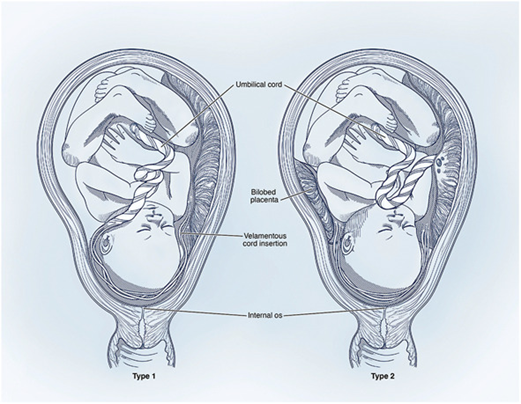
Fig. 1. Type 1 and 2 vasa previa
Oyelese. Vasa Previa. Obstet Gynecol 2023. Illustration by Alex Webber (http://www.dnaillustrations.com).
Signs and symptoms of vasa previa
Vasa previa does not always show symptoms; most people will only get diagnosed through their routine pregnancy ultrasound. However, pregnant people affected by vasa previa might experience symptoms such as:
During pregnancy:
- Vaginal bleeding: occurring from your second trimester of pregnancy onwards, the blood is darker than period blood because it is the foetus’s blood and not the pregnant person's.
- Decreased fetal movement
- Abdominal pain
During active labour:
- Change in foetus heart rate
- ruptures of membranes
- pale newborn1,2
Risk Factors and causes
There are different risk factors associated with vasa previa. You are more at risk of contracting vasa previa if you are pregnant with multiples. Similarly, you are more at risk if you have a velamentous cord insertion or a placenta with accessory lobes. Placenta previa occurs when the placenta partially or fully covers the cervix, and a low-lying placenta during pregnancy is an additional risk factor that elevates the likelihood of vasa previa. If previously you had uterine surgery, your chances of developing vasa previa are higher, as surgical procedures can lead to uterine scarring. Finally, studies have shown that assisted reproductive pregnancies, such as in vitro fertilisation are associated with a significantly elevated risk of vasa previa. Approximately 26% of vasa previa cases occur in pregnancies conceived through assisted reproductive technology.2
Diagnosis and Screening
Vasa previa is most commonly picked up by prenatal ultrasound imaging. The medical team may use colour Doppler for the ultrasound to be more precise. Colour doppler is an imaging technique that will use high-frequency sound waves to observe blood flow in the placenta and the umbilical cord, which will help in diagnosing vasa previa and understanding how the vessels are arranged.3
Further tests, such as MRI can also be completed if the ultrasound is not conclusive to detect vasa previa.
Management and treatment for vasa previa
There is no way to directly treat vasa previa. The most common management technique when diagnosed with vasa previa is to schedule a caesarean section (C-section) delivery at around 36 weeks of gestation. This will allow the best outcomes for patients with vasa previa, as it avoids the pregnant person going into labour and reduces the chance of membrane rupture. Along with this procedure, future pregnant persons should avoid intense physical activities and sexual intercourse.
Your doctor might recommend corticosteroids (anti-inflammatory medicine) to facilitate fetal lung development in anticipation of a C-section delivery. Additionally, your doctor might arrange frequent (typically twice a week) CTG monitoring, which will evaluate the fetal heart rate. Finally, you might be taken into the hospital for 24/7 medical supervision at the end of your pregnancy to be closely monitored during the third trimester of pregnancy.1,2,4
Non-diagnosed vasa previa
On the other hand, if vasa previa is not diagnosed before labour, the treatment when the fetal vessels rupture is going to be blood transfusion and aggressive newborn resuscitation by the medical team. Nonetheless, despite management, a substantial risk of neonatal hypovolemic and hypoxemic injury persists, along with potential neurodevelopmental impairment for the newborn.2
New therapies under investigation
In order to cure vasa previa, new therapies have been explored. A procedure called fetoscopic laser ablation is believed to hold promise as a cure for vasa previa. The medical procedure involves sealing the unprotected fetal vessels with a laser to prevent vessel rupture during labour. This procedure would be done at around 32 weeks pregnant and would allow you not to get hospitalised and have a vaginal full-term labour. The problem with this procedure is that undergoing Fetoscopic laser ablation could lead to infection, membrane rupture, premature delivery, fetal blood loss and potentially unknown long-term effects on placental health. Due to the risks, this method is still under investigation and cannot be performed in the hospital as of yet.2
Summary
In summary, Vasa Previa is a rare but serious pregnancy complication characterised by fetal blood vessels crossing the cervix. When these vessels rupture during labour, it can lead to fetal blood loss and, in severe cases, fetal death. This condition, though rare with an incidence rate of approximately 1 in 2,500 deliveries, is more prevalent in pregnancies with multiples and in vitro fertilisation (IVF) cases.
Early diagnosis through routine pregnancy ultrasound is crucial. Management primarily involves scheduling a C-section around 36 weeks of gestation, along with close monitoring and, in some cases, corticosteroid treatment to support fetal lung development.
For non-diagnosed cases during labour, treatment includes blood transfusion and newborn resuscitation, but risks and complications remain. New therapies, such as Fetoscopic Laser Ablation, are under investigation to address Vasa Previa, although they come with potential risks and uncertainties. To address Vasa Previa, awareness, early diagnosis, and prompt medical attention remain vital to the safety of the pregnant person and the baby's well-being.
References
- 1.Vasa Previa: Causes. Symptoms, Management & Treatment. Cleveland Clinic [Internet]. [cited 2023 Oct 7]. Available from: https://my.clevelandclinic.org/health/diseases/23465-vasa-previa.
- 2.Oyelese, Y., Javinani, A., & Shamshirsaz, A. A. (2023). Vasa Previa. Obstetrics & Gynecology, 142(3), 503–518. https://doi.org/10.1097/aog.0000000000005287
- Vasa Previa: Causes. Symptoms, Management & Treatment. Cleveland Clinic . [cited 2023 Oct 7]. Available from: https://my.clevelandclinic.org/health/diseases/23465-vasa-previa.
- 3. Shah, Aalap, and Abid Irshad. "Sonography Doppler Flow Imaging Instrumentation." StatPearls. StatPearls Publishing, 2023.
- 4. Vasa Previa Fact Sheet. [cited 2023 Oct 7]. Available from: https://vasaprevia.com/Vasa-Previa-Fact-Sheet#:~:text=Vasa%20 previa%20must%20be%20 present,velamentous%20insertion%20of%20the%20cord%2C.


